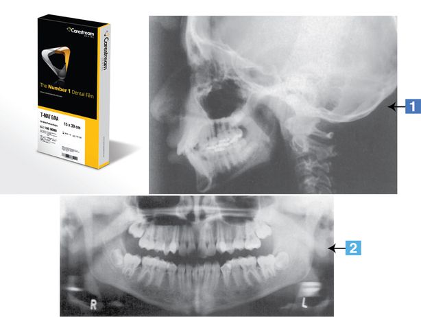
Dental radiography equipment
 Instrument
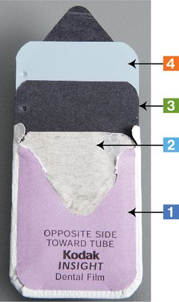
Intraoral dental film


Practice note 
 Follow standard precautions and cross-contamination protocol when exposing and processing film for developing. Outer packet and black paper may be disposed of in the garbage. Correct disposal of lead foil must be checked within your state. In some states, lead foil is considered a hazardous waste and must be collected and disposed of properly. For proper recycling protocol, refer to Department of Environmental Health regulations in the state where you practice.
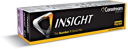
Photos courtesy Carestream Health Inc., Rochester, NY.
 Instrument
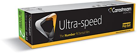
Package of dental film


Practice notes 
 Follow standard precautions and cross-contamination protocol when processing film for developing. Outer packet and black paper may be disposed of in the garbage. Correct disposal of lead foil must be checked within your state. In some states, lead foil is considered a hazardous waste and must be collected and disposed of properly. For proper recycling protocol, refer to Department of Environmental Health regulations in the state where you practice.
 Instrument
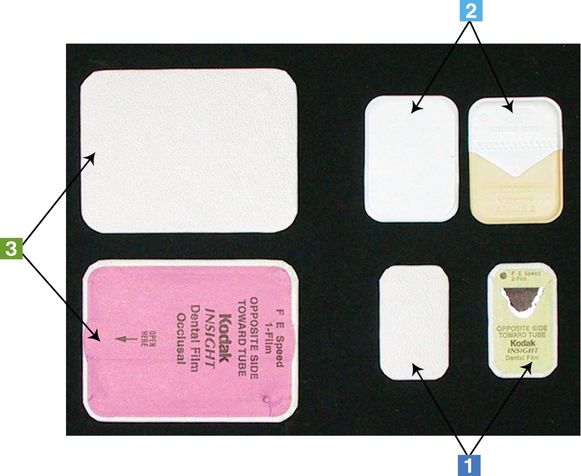
Intraoral dental film—various sizes


Practice note 
 Follow standard precautions and cross-contamination protocol when exposing and processing film for developing. Outer packet and black paper may be disposed of in the garbage. Correct disposal of lead foil must be checked within your state. In some states, lead foil is considered a hazardous waste and must be collected and disposed of properly. For proper recycling protocol, refer to Department of Environmental Health regulations in the state where you practice.
Product photo courtesy Carestream Health Inc., Rochester, NY.
 Instrument


 Follow standard precautions and cross-contamination protocol when exposing and processing film for developing.
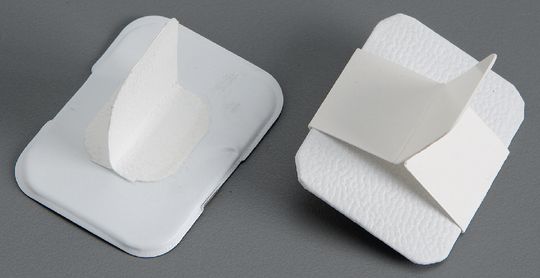
 Instrument
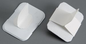
Bite-wing tabs


Practice notes 
 Follow standard precautions and cross-contamination protocol when exposing and processing film. Outer packet, black paper, and Bite-Wing Tabs may be disposed of in the garbage. Correct disposal of lead foil must be checked within your state. In some states, lead foil is considered a hazardous waste and must be collected and disposed of properly. For proper recycling protocol, refer to Department of Environmental Health regulations in the state where you practice.
 Instrument
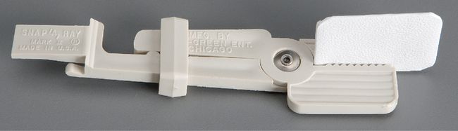
Film holder—periapical (eezee-grip)


Practice notes 
 EeZee-Grip Film Holder must be cleaned, bagged individually, and then sterilized. A chemical/steam indicator device should be included in the wrapping.
Photos courtesy DENTSPLY Rinn, Elgin, IL.
 Instrument


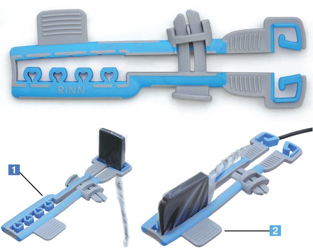
 Follow standard precautions and cross-contamination protocol when taking digital images. Holder must be cleaned, bagged individually, and then sterilized. A chemical/steam indicator device should be included in the wrapping.
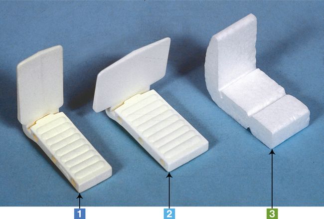
 Instrument


 Plastic Film Holders must be cleaned, bagged individually, and then sterilized. A chemical/steam indicator device should be included in the wrapping. Disposable Styrofoam holder may be disposed of in garbage.
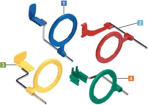
 Instrument


Stay updated, free dental videos. Join our Telegram channel
VIDEdental - Online dental courses


