Introduction
In preorthodontic children with Class II malocclusion and horizontal maxillary overjet, cervical column morphology was examined and related to craniofacial morphology and head posture for the first time.
Methods
Two hundred thirteen children (aged 7-15 years) with a horizontal maxillary overjet of more than 6 mm were divided into 2 groups of skeletal and dentoalveolar overjets. The skeletal overjet group comprised 99 patients (43 girls, 56 boys). The dentoalveolar overjet group comprised 114 subjects (58 girls, 56 boys). Visual assessments of the cervical column and measurements of craniofacial morphology and head posture were made on profile radiographs.
Results
Deviations in the cervical vertebral column morphology occurred significantly more often in the skeletal overjet group (28%) compared with the dentoalveolar overjet group (17%) ( P <0.05). Fusion anomalies were associated with a large sagittal jaw relationship, retrognathia of the jaws, large inclination of the jaws, and extended head posture ( P <0.05 and 0.01, respectively). Furthermore, a partial cleft was significantly associated with a large cranial base angle ( P <0.01).
Conclusions
New associations were found between cervical column morphology, craniofacial morphology, and head posture in preorthodontic children with horizontal maxillary overjet. These findings are considered important for diagnostics and thus for a more accurate treatment plan of these patients.
Deviations of the cervical column morphology occur in healthy subjects with neutral occlusion and normal craniofacial morphology as well as in patients with craniofacial syndromes, deviating craniofacial morphology, and severe malocclusion traits. A recent study found that fusions between the upper cervical vertebrae (C2 and C3) occurred in 14.3% of healthy subjects. Fusions of the upper cervical column within that range are thus considered normal.
Previous studies have found an association between malformations of the upper cervical vertebrae and patients with cleft lip and palate. Recently, an association was also found between malformation of the upper cervical vertebrae not only in patients with condylar hypoplasia, but also in adult orthodontic surgical patients with skeletal deep bite, skeletal mandibular overjet, skeletal horizontal overjet, and skeletal open bite. These studies showed that cervical column deviations occurred in 72.7% of the condylar-hypoplasia group, 41.5% of the deep-bite group, 61.4% of the mandibular-overjet group, 52.9% of the horizontal-overjet group, and 42.1% of the open-bite group. Deviations occurred significantly more often in all 5 patient groups compared with the control group. This indicates that morphologic deviations of the upper cervical vertebrae are not only associated with malformation of the jaws but also with craniofacial morphology and occlusion.
A previous study of adults found that fusion between C2 and C3 was significantly associated with posture of the head and neck. In this study, the cervical vertebral column was approximately 5° more curved, and the inclination of the upper cervical spine was 8° more backward in adults with fusion.
Accordingly, it is relevant to focus on similar associations between cervical column morphology, craniofacial morphology, and posture of the head and neck in preorthodontic children. To our knowledge, no studies have been performed so far on cervical column morphology in relation to craniofacial morphology and head posture in preorthodontic children with Class II malocclusion and horizontal maxillary overjet.
Our aims in this study were to (1) describe the morphology of the cervical column in children with skeletal horizontal maxillary overjet and dentoalveolar horizontal maxillary overjet, (2) compare the morphology of the cervical column in a group of children with skeletal horizontal maxillary overjet (the skeletal overjet group) with a group of children with dentoalveolar horizontal maxillary overjet (the dentoalveolar overjet group), and (3) analyze associations between the morphology of the cervical column, craniofacial dimensions, and head posture in both groups together.
Material and methods
Two hundred thirteen profile radiographs were systematically selected according to the inclusion criteria mentioned below from patients registered between 1988 and 1997 at the orthodontic clinic of the municipal dental service of Farum, Denmark, and divided into 2 groups according to type of overjet: skeletal and dentoalveolar.
The skeletal overjet group comprised 99 patients, 43 girls (ages, 8-14 years; mean, 10.2 years) and 56 boys (ages, 7-15 years; mean, 10.0 years). The inclusion criteria for the skeletal horizontal maxillary overjet group were (1) no prior orthodontic treatment, (2) a skeletal horizontal maxillary overjet of more than 6 mm (the sagittal jaw relationship larger than 1 SD according to the standard described by Björk, assessed on the lateral radiograph of each subject), (3) no craniofacial anomalies or systemic muscle or joint disorders, and (4) accessibility of a profile radiograph before orthodontic treatment with the first 5 cervical vertebrae visible. The sagittal jaw relationship ranged between 4.5° and 14.0° (mean, 6.3°), the horizontal overjet was 6.0 to 12.7 mm (mean, 8.3 mm), the vertical jaw relationship was 14.4° to 39.2° (mean, 27.9°), and the vertical overbite was between −3.4 and 7.6 mm (mean, 2.5 mm) ( Table I ).
| Variable | Skeletal (n = 99) | Dentoalveolar (n = 114) | Group | Sex | ||
|---|---|---|---|---|---|---|
| Mean | SD | Mean | SD | P | P | |
| Cranial base angle (°) | ||||||
| N-S-Ba | 133.2 | 4.9 | 132.7 | 5.6 | NS | NS |
| Sagittal dimensions (°) | ||||||
| ss-n-pg | 6.3 | 1.6 | 2.5 | 1.5 | ‡ | NS |
| s-n-ss | 81.7 | 4.1 | 79.7 | 4.0 | ‡ | NS |
| S-N-Pg | 75.4 | 4.1 | 77.2 | 4.1 | † | NS |
| Vertical dimensions (°) | ||||||
| NL-ML | 27.9 | 4.8 | 24.1 | 5.5 | ‡ | NS |
| NSL-NL | 7.2 | 3.7 | 6.8 | 3.2 | NS | |
| NSL-ML | 35.0 | 6.0 | 30.9 | 5.9 | ‡ | NS |
| Incisor relations (mm) | ||||||
| Overjet | 8.3 | 1.5 | 7.9 | 1.4 | * | * 2 |
| Overbite | 2.5 | 2.3 | 3.1 | 1.9 | * | † 2 |
| Head posture (°) | ||||||
| NSL/VER | 101.2 | 5.6 | 100.5 | 5.4 | NS | NS |
| NL/VER | 94.0 | 4.6 | 93.7 | 4.3 | NS | NS |
| NSL/OPT | 96.2 | 8.3 | 96.7 | 8.5 | NS | NS |
| NL/OPT | 89.0 | 7.7 | 89.9 | 7.8 | NS | NS |
| NSL/CVT | 100.8 | 8.1 | 101.3 | 8.5 | NS | † 1 |
| NL/CVT | 93.6 | 7.2 | 94.5 | 7.6 | NS | ‡ 1 |
| OPT/HOR | 95.0 | 7.7 | 93.9 | 7.5 | NS | * 2 |
| CVT/HOR | 90.4 | 6.9 | 89.2 | 7.1 | NS | ‡ 2 |
| OPT/CVT | 4.6 | 2.9 | 4.6 | 3.0 | NS | † 1 |
The dentolveolar overjet group comprised 114 subjects, 58 girls (ages, 7-15 years; mean, 10.7 years) and 56 boys (ages, 7-15 years; mean, 10.7 years). The inclusion criteria for the dentoalveolar overjet group were (1) no prior orthodontic treatment, (2) a dentoalveolar horizontal maxillary overjet of more than 6 mm (the sagittal jaw relationship smaller than 1 SD according to the standard described by Björk, assessed on the lateral radiograph of each subject), (3) no craniofacial anomalies or systemic muscle or joint disorders, and (4) accessibility of a profile radiograph before orthodontic treatment with the 5 first cervical vertebrae visible. The sagittal jaw relationship ranged between 1.6° and 4.4° (mean, 2.5°), the horizontal overjet was 6.0 to 12.7 mm (mean, 7.9 mm), the vertical jaw relationship was 9.2° to 37.6° (mean, 24.1°), and the vertical overbite was between −3.0 and 6.6 mm (mean, 3.3 mm) ( Table I ).
The differences in mean values for the craniofacial dimensions between the 2 groups and sexes are presented in Table I .
The morphology of the cervical column was evaluated by visual inspection of the first 5 cervical vertebrae as they are normally seen on a standardized lateral skull radiograph. Characteristics of the cervical column were classified according to the method of Sandham and divided into 2 categories.
- 1.
Posterior arch deficiency, defined as partial cleft and dehiscence. Partial cleft is defined as failure to fuse of the posterior part of the neural arch ( Fig 1 ), and dehiscence is defined as failure to develop of a part of a vertebral unit.
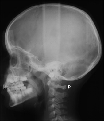
Fig 1 A profile radiograph illustrating a partial cleft of the posterior part of the neural arch of the atlas ( P ). - 2.
Fusion anomalies, defined as fusion, block fusion, and occipitalization. Fusion is defined as fusion of 1 unit with another at the vertebral bodies, articulation facets, neural arch, or transverse processes ( Fig 2 ). Occipitalization is defined as assimilation, either partially or completely, of the atlas (C1) with the occipital bone ( Fig 3 ). The definition of block fusion was modified according to the method of Sonnesen and Kjær and defined as fusion of more than 2 units at the vertebral bodies, articulation facets, neural arch, or transverse processes.

Fig 2 A profile radiograph illustrating fusion of the C2 and C3 vertebrae ( F ).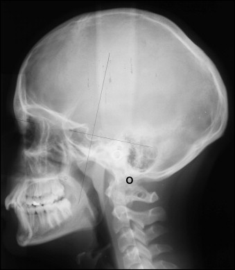
Fig 3 A profile radiograph illustrating occipitalization between the C1 vertebra and the occipital bone ( O ).
Only anomalies verified on the later profile radiographs were registered as anomalies of the cervical vertebral column.
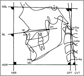
The profile radiographs were taken with the teeth in occlusion and in the standardized head posture, the mirror position, as described by Siersbæk-Nielsen and Solow. The radiographs were taken in a cephalostat with a film-to-focus distance of 180 cm and a film-to-median plane distance of 10 cm. No correction was made for the constant linear enlargement of 5.6%. The reference points were marked and digitized directly on the profile radiographs by using a Tiops 2000 digitizer (version 2.7.0, Total Interactive Orthodontics Planning System, TIOPS, Denmark) and analyzed digitally by using Tiops 2005 (version 2.12.4) ( Fig 4 ). Eighteen variables representing the cranial base angle, the vertical and the sagittal craniofacial dimensions, and the head posture were calculated. A list of the variables is given in Table I .
Reliability of the visual assessment of the morphologic characteristics of the cervical vertebral units was determined by interobserver examination, which showed very good agreement (0.82) as assessed by the kappa coefficient.
Reliability of the variables describing the cranial base, the vertical and sagittal craniofacial dimensions, and the head posture was assessed by remeasuring 20 lateral radiographs selected at random from the previously evaluated radiographs. The radiographs were digitized again, and paired t tests found no significant differences between the 2 sets of recordings. The method errors calculated by Dahlberg’s formula ranged from 0.09° to 0.69°, and the Houston reliability coefficients were from 0.99 to 1.00. The reliability was within the same range as for traditional film-based radiographs.
Statistical analysis
For the craniofacial dimensions, the effect of age was assessed by linear regression analysis and the morphologic characteristics of the cervical column by logistic regression analysis. Differences in the means of the craniofacial dimensions between sexes and groups were assessed by unpaired t tests. Differences in morphologic characteristics of the cervical column between sexes and groups were assessed by the Fisher exact test. Associations between morphology of the cervical column and each craniofacial dimension were expressed in terms of the Nagelkerke logistic regression correlation coefficients (R 2 ) (R = √R2
R 2
) and tested for the possible effect of age and sex by multiple logistic regression analyses. The results from the tests were considered to be significant at P values below 0.05. The statistical analyses were performed by using SPSS software (version 13.00, SPSS, Chicago, Ill).
Stay updated, free dental videos. Join our Telegram channel
VIDEdental - Online dental courses


