Fig. 5.1
(a, b) The patient arrived at the dental office with a chief complaint of recurrent abscesses in the past year. The left mandibular molar was tender to percussion and palpation, and cl II mobility was also noted. An 8 mm probing defect was recorded in the mesiobuccal area and 5 mm in the midbuccal. The periapical radiograph (a) showed a previously treated mandibular molar, gutta-percha–filled root canals, a Dentatus dowel in the distal root, an amalgam dowel in the mesial, and full coverage with a crown. There was bone loss in the bifurcation and at the coronal half of the distal root. The mesial root was surrounded by a radiolucent lesion. A VRF was suspected and the tooth extracted. The typical buccolingual fracture can be seen in Fig. 5.1b
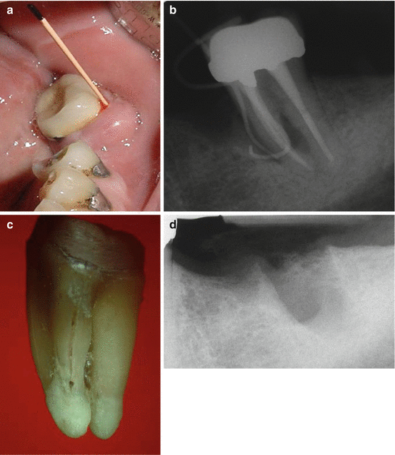
Fig. 5.2
(a–d) The patient arrived at the dental office with “soreness on biting for the last couple of months and a swelling in the area about a year ago.” The first mandibular molar was extracted 5 years earlier but only the second molar endodontically treated, restored with amalgam dowels in the coronal 3 mm of the two roots and a crown (a). Clinical examination revealed sensitivity to percussion and two 8 mm probing defects both in the buccal and lingual aspects. The periapical radiograph (b) shows two gutta-percha tracing cones at the bone resorption area in the coronal two-thirds of the mesial root. A VRF diagnosis was made and the tooth extracted. In the extracted tooth (c), the fracture in the coronal two-thirds of the mesial root can be seen. A periapical radiograph of the extracted site (d) shows the amount of bone loss in the bone as a result of the continuous inflammatory process in the area (Courtesy Prof. J. Nissan)
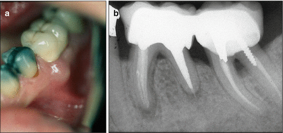
Fig. 5.3
(a, b) A patient with severe periodontal problems who was evaluated for periodontal surgery arrived at the office with a small swelling adjacent to the mesial root area (a). The mandibular left first molar was endodotically treated 7 years previously and restored afterward. Probing depth around the tooth was 5 mm in the midbuccal and 4 mm in the mesiobuccal area. The radiographic appearance of the radiolucency around the mesial root which most likely was of an endodontic origin (b) and swelling in the gingivae adjacent to the mesial root pointed to a VRF. However, in this periodontally involved patient, the final diagnosis was an acute apical abscess (Courtesy Dr. J Halpern)
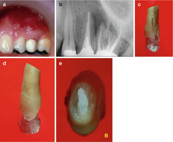
Fig. 5.4
(a–e) A patient arrived at the dental office with a complaint of “draining pus from the gum” in the upper jaw. Clinical examination revealed a veneer crown in the second premolar that was endodontically treated and restored at the same time with the maxillary molar adjacent to it (a). A highly located sinus tract was noted, but the probing numbers were between normal limits. Endodontic treatment appeared adequate in the radiograph (b), but a large radiolucent area can be seen laterally in the distal aspect of the premolar tooth involving the mesiobuccal root of the maxillary molar. Poor outcome of the endodontic treatment either in the maxillary premolar (chronic apical abscess) or the mesiobuccal root of the molar led to the treatment plan of endodontic surgery either in the premolar or the mesiobuccal root or both. Endodontic surgery was performed in the maxillary premolar with IRM as a retrograde filling material. At 12 months post-op, the patient returned with an acute abscess, and the premolar was extracted. A VRF was noted in the buccal aspect (c), but it was of the incomplete type since it was not seen in the buccal aspect (d). In the apical view (e), the incomplete VRF can be seen in the palatal aspect but not in the buccal one. Most likely, the incomplete VRF on the palatal aspect was not noticed during surgery
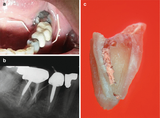
Fig. 5.5
(a–c) The mesial root of the mandibular molar showing two gutta-percha tracings from the buccal and lingual sinus tracts (a). Radiograph (b) shows the wide lateral radiolucency on the mesial root aspect. Also, there is radiolucency in the bifurcation. After VRF diagnosis and extraction, the crown was removed, and one part of the mesial root can be seen with some of the gutta-percha filling (c) (Courtesy Dr. A. Aronovich)
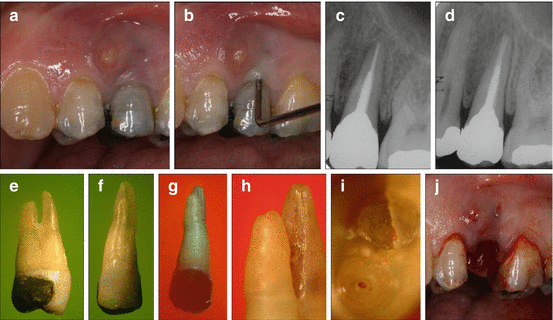
Fig. 5.6
(a–j) A patient arrived at the dental office with a complaint of a “lump in the gum” for several months (a). From the case history, it was revealed that the maxillary second premolar was endodontically treated 3 years earlier and Dentatus and a large amalgam restoration followed thereafter. Clinical evaluation revealed a highly located sinus tract (a, b), and a narrow midbuccal 7 mm probing defect was noted (b). A small isolated lateral radiolucency on the mesial aspect was noted along the root (c, d). A VRF diagnosis was made and the tooth extracted. It was a bifurcated maxillary premolar (e) of which only the buccal root was fractured (f) but not the lingual one (g). A closer examination of the two roots shows the typical depression on the bifurcation aspect of the buccal root (R) along with the apicocoronal VRF. A closer look at the two apices (i) the complete buccolingual VRF on the buccal root. After tooth extraction, the typical bony dehiscence, which faced the fracture in the buccal root, is reflected as triangular flabby attached gingivae (j) (Courtesy Dr. E. Venezia)
Stay updated, free dental videos. Join our Telegram channel

VIDEdental - Online dental courses


