(b) 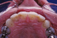
Table 8.1.1 Clinical signs of external cervical resorption
|
A periapical radiograph of the tooth revealed a radiolucency in the cervical region of the tooth; this lesion was 5–6 mm wide (Figure 8.1.2a). A second radiograph was taken with a 10° change in the horizontal plane (horizontal parallax) of the X-ray tube head (Figure 8.1.2b); the radiolucency on the radiograph moved in the opposite direction to the tube shift, in relation to the root canal. The outline of the root canal could be traced through the lesion on each radiograph, and there was no periapical radiolucency associated with the tooth on either radiograph (Table 8.1.2). A limited cone beam computed tomography (CBCT) scan was taken to assess the true nature of the lesion (Figure 8.1.3).
Figure 8.1.2 (a) Periapical radiograph of the 21, (b) a second periapical radiograph taken of the 21 with a shift in the direction of the X-ray tube to the left. The resorptive lesion has moved to the right on the radiograph in 8.1.2b as compared to 8.1.2a confirming that the lesion is confined to the labial aspect of the tooth. The outline of the root canal can be traced through the lesion in both radiographs.
(a) 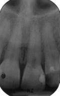 (b)
(b) 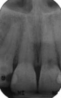
Table 8.1.2 Radiological signs of external cervical resorption
|
Diagnosis and treatment planning
What was the diagnosis?
The diagnosis was external cervical root resorption (ECR).
Why were two radiographs taken?
Internal root resorption and external cervical root resorption can have similar appearances on conventional radiographs, and may be confused with each other. Two conventional radiographic features are of particular help in differentiating between the conditions. These are:
- The presence of the root canal outline through the lesion.
- The movement of the lesion on parallax radiographs.
In the case of internal root resorption, the outline of the root canal cannot usually be traced through the lesion on the radiograph. This is because an internal root resorption defect originates within, and is therefore continuous with the root canal space. On the other hand, an external cervical root resorption lesion begins on the external surface of the root, progresses inwards, and does not communicate with the root canal until it is very advanced and perforates the root canal wall. As a result, it is generally possible to trace the outline of the intact root canal walls through the external cervical root resorption lesion on a radiograph.
A second radiograph taken with a change in angle of the X-ray beam will result in a change in position of an external cervical root resorption lesion, whilst an internal resorptive lesion will not change position on parallax radiographs. In addition, parallax radiographs will allow the clinician to determine the position of an external cervical root resorption lesion using the ‘SLOB’ (Same Lingual Opposite Buccal) rule. If the external cervical root resorption lesion moves in the same direction as the change in angle of the X-ray beam, then the lesion is lingually or palatally located. If it moves in the opposite direction, it is buccally located.
What are the limitations of conventional radiographs for assessing complex anatomical problems (i.e. root resorption lesions)?
Stay updated, free dental videos. Join our Telegram channel
VIDEdental - Online dental courses


