(b) 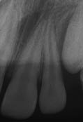 (c)
(c) 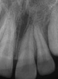
Diagnosis and treatment planning
The diagnoses (Tables 7.1.1 and 7.1.2) were:
- Upper left central incisor (21) – Tooth concussion.
- Upper left lateral incisor (22) – Subluxation.
Table 7.1.1 Clinical and radiographic features of concussion injuries
|
Table 7.1.2 Clinical and radiographic features of subluxation injuries
|
The treatment plan was to provide symptomatic treatment and monitor the pulpal responses to the injury (Table 7.1.3).
Table 7.1.3 Treatment of concussion and subluxation injuries
|
What are the types of luxation injuries?
There are five types of luxation injuries:
Concussion
The tooth is sensitive to biting and percussion, but there is no increased mobility and the tooth has not been displaced. The impact of injury has affected the PDL, and there can be some slight damage to the neurovascular supply to the pulp.
Subluxation
In addition to increased sensitivity to biting and percussion, there is also increased mobility. The tooth, however, has not been displaced. There is damage to the PDL and the alveolar socket; in addition, there may be injury to the neurovascular supply to the pulp.
Extrusive luxation
The tooth has been displaced out of the socket, often severing the neurovascular supply to the pulp. The tooth is very mobile, and there can be a concomitant alveolar fracture. Sensibility testing is usually negative.
Lateral luxation
The tooth has been displaced laterally into the adjacent bony support. Typically, the crown has been displaced palatally/lingually, and the root has shifted toward the labial cortical plate. The tooth is usually very firmly embedded in its new position and has an ankylotic sound when percussed.
Intrusive luxation
Stay updated, free dental videos. Join our Telegram channel
VIDEdental - Online dental courses


