4: Initial Assessment and Intensive Care of the Trauma Patient

Initial Assessment and Intensive Care of the Trauma Patient

Outline

Patient Transport

History of Medical Transportation

Prior to the 1600s, several methods of moving the debilitated were in use around the world. In what would become the United States, Native Americans used the travois (Fig. 4-1), which was essentially a stretcher affixed at one end to a horse or large dog to pull as a conveyance for material or disabled people. In Egypt, camel stretchers called panniers, which were used in many regions around the world with other beasts of burden such as mules, were the method of choice, even through the Napoleonic period, to move the nonambulatory.

In the fifteenth century, King Ferdinand and Queen Isabella of Spain were instrumental in the deployment of ambulancia (mobile field hospitals) to provide rapid medical aid to their soldiers. In January 1777, George Washington instructed the army’s Hospital Department Director General, Dr. John Cochran, to consult with another well-known physician of the day and his successor, Dr. William Shippen, to reorganize the medical system, including the flying hospitals, to accompany his armies in the field. Flying hospitals were equivalent to today’s MASH units (mobile army surgical hospitals). They were semitransient field hospitals that were moved with the regiment or divisional army, providing immediate care to those wounded after a battle and to those who were ill.

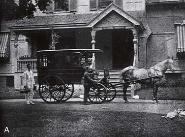
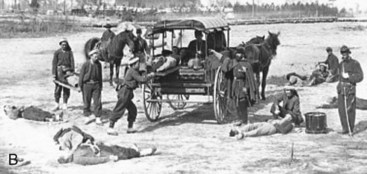
The next significant development of U.S. emergency medical services (EMS) and medical transport took place during the Civil War (1861-1865). The Union Medical Department implemented the use of committed, customized horse-drawn wagons as ambulances (Fig. 4-2A) as well as stretcher litters and pack animal cacolets (see Fig. 4-2B). In addition, a dedicated group of stretcher bearers and ambulance wagon attendants and drivers was formed. They received specialized training by the medical department and a tiered transport system was developed. On March 11, 1864, President Lincoln signed a law that was passed by Congress, “An Act to Establish a Uniform System of Ambulances in the Armies of the United States.” This legislation established a standardized system of ambulance service throughout the military. The law also mandated the use of special uniforms for the ambulance corps and special signs for the ambulances. Regulations issued during the war by both sides, and incorporated into this law, also conventionalized specific insignia and signage for the recognition of ambulances and hospitals.

In addition to litters, cacolets, and wagons, trains and boats were also used as medical transportation vehicles on a regular basis. Ambulance wagons took the wounded and sick from field hospitals to trains or hospital ships instead of directly to the general hospital. This would occur if the general hospital was too far to allow for reasonable transport by wagon, train, or ship. Thus, a stratified system of medical transport and transport vehicles developed.

The first civilian-manufactured ambulance in the United States, a specialized medical transport vehicle, was built in 1890 by the Hess-Eisenhardt Company of Cincinnati, Ohio. It was a horse-drawn wagon specifically designed to move the incapacitated in need of medical care. Shortly thereafter, the first motorized ambulance was made in Chicago and donated to the Michael Reese Hospital by five local businessmen in 1899. This was quickly followed by St. Vincent’s Hospital of New York, which began operating an automobile ambulance in 1900. In 1910, the first known aircraft ambulance, a plane modified to carry a patient lying down, was built in North Carolina and tested in Florida. It failed shortly following take-off and crashed after flying only 400 yards in Fort Barrancas, Florida. By 1929, the U.S. Army Air Corps had been organized and designed three planes to perform as ambulances. They were built and equipped to carry two patients on stretchers, a pilot, and an attendant.

World War II, the Korean Conflict, and the Vietnam War all brought advances in trauma medicine to the military, which were then used in the civilian sector. Some improvements in military EMS organization and operations occurred; the most noteworthy was the use of helicopters to retrieve critically injured patients rapidly from the battlefield, transporting them to field hospitals (MASH units). Reducing the time from injury to surgical intervention, with on-scene advanced medical treatment such as the administration of IV fluids by nonphysicians, marked a milestone in the progression of clinical care.

A convergence of several landmark events occurred in the mid-1960s that altered the foundations of EMS in the United States, commencing a new era of modernity and organization. For decades, a crescendo had been building regarding the carnage experienced from automobile accidents on the nation’s highways. Finally, in 1961, President John F. Kennedy noted that traffic accidents were one of the greatest of the nation’s public health problems. Instantly, a new focus was established on the need for emergency medical aid for motor vehicle accident (MVA) victims.

Several crucial events took place during President Johnson’s administration. First, in 1965, Medicare was created by an act of Congress. In the original legislation, ambulance transportation was recognized as a covered beneficiary service. In so doing, the federal government established a long-term funding mechanism for EMS and medical transportation. The introduction and propagation of prehospital medical care, especially advanced clinical treatment, would not wait for government regulations. Often with no legislation regarding their activities, U.S. physicians were spearheading the use of medications, defibrillators, and other advanced medical treatment modalities in the field, at the scene of incipient need. Most of these services started with a single-minded focus of cardiac emergencies, but rapidly expanded into treating other medically urgent conditions. The emphasis was shifting from the rapid recovery and transport of victims to the rapid response of specialized personnel and apparatus, and the stabilization of patients before movement to a hospital.

The first advanced life support ambulance in the United States was used by St. Vincent’s Hospital in New York. The first nonphysician, mobile, advanced medical treatment service in America began in Miami in 1968. Dr. Eugene Nagel blended the training of surrogate quasiphysicians with radiotechnology to devise the concept of a paramedic using telemetry communication to receive real-time medical commands from a physician at the hospital. This service was followed shortly thereafter by similar programs in Columbus, Ohio, Jacksonville, Florida, Seattle, and Los Angeles. By the end of the 1970s, EMS was firmly established in the medical infrastructure of the United States as its own discipline, with its own science. During the next several decades, it would become more sophisticated, evolving into an industry.

Types of Transport

Helicopter

Helicopters offer several advantages over other transport vehicles (Fig. 4-3). They can travel at speeds of 120 to 180 mph, allowing for transport times to be up to 75% shorter when compared with ground transport. They can avoid traffic delays and ground obstacles and fly into locations inaccessible by other modes of patient transport. Helicopters have landing requirements, which is a disadvantage when compared with ground transport, but they can access more regions than fixed-wing transport. Helicopter cabins are not pressurized and, as a result, patients being transported are at some risk for barotrauma. Another disadvantage is that in most transport programs, helicopters are only permitted to fly under visual flight rules. Hence, weather conditions can limit this operation. However, some programs are now implementing instrument flight rules, which give greater flexibility to flying in less than ideal weather.5

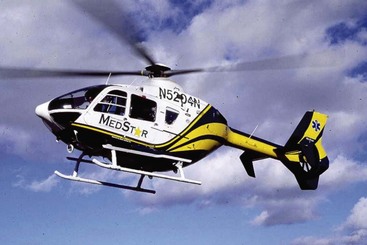
The widespread use of helicopters for patient transport has led to increased accident rates (three times higher) when compared with general helicopter aviation.6 Another study has demonstrated a lower accident rate among busier flight programs and programs that implement instrument flight rules.7 Helicopter ambulances are best used when hospital ground transport time is expected to be longer than 35 minutes or when ground transport is not a viable option. Helicopter transport of critically injured patients from remote areas may be lifesaving; however, there is a large potential for its misuse. The annual cost of helicopter transport service can be at least $1 million for an institution. For effective use of this transport modality, it is generally recommended that helicopter transport should be integrated into the regional EMS system, staffed to provide advanced life support, and used based on medical need.

Severely injured patients transported by helicopter from the scene of an accident are more likely to survive than patients brought to trauma centers by ground ambulance, according to a recent study. This study was the first to examine the role of helicopter transport on a national level and includes the largest number of helicopter transport patients in a single analysis. The finding that helicopter transport positively affects patient survival comes amid an ongoing debate surrounding the role of helicopter transport in civilian trauma care in the United States, with advocates citing the benefits of fast transport times and critics pointing to safety, use, and cost concerns. The new national data show that patients selected for helicopter transport to trauma centers are more severely injured, come from greater distances, and require more hospital resources, including admission to the intensive care unit (ICU), use of a ventilator to assist breathing, and urgent surgery compared with patients transported by ground ambulance. Despite this, helicopter transport patients are more likely than ground transport patients to survive and be sent home following treatment.

Air medical transport is a valuable resource that can make trauma center care more accessible to patients who would not otherwise be able to reach these centers. The study included patients transported from the scene of an injury to a trauma center by helicopter or ground transportation in 2007. The team used the National Trauma Databank to identify 258,387 patients; 16% were transported by helicopter and 84% were transported by ground. The helicoptertransport patients were younger, more likely to be male, and more likely to be victims of MVAs or falls compared with ground transport patients. Overall, almost 50% of the helicopter transport patients were admitted to the ICU, 20% required assistance in breathing for an average of 1 week, and almost 20% required surgery. Even though they were more debilitated when they arrived at the hospital, they ultimately fared better than those transported by ground.

Although the study has shown that air transport does make a difference in patient outcomes, there are no data available to explain why patients transported by helicopter do better than those transported by ground. The authors assumed that the speed of transport—helicopters are capable of higher speeds over longer distances, regardless of terrain—and the ability of air medical crews to provide therapies and use treatment modailities not universally available to ground unit crews are the main drivers of positive patient outcomes. However, the study has some limitations. It is not possible to evaluate the many factors that drive the decision to transport a patient by helicopter in all cases. In addition, the general nature of the data set limits specific conclusions that could be applied to any individual trauma system.

Helicopter transport has been an integral component of trauma care in the United States since the 1970s, mainly because of the military’s experience in transporting sick or injured soldiers during wartime. The availability of helicopters in the civilian setting has been credited with improving trauma center access for a significant percentage of the population.

Fixed-Wing Air Ambulance

This mode of patient transport is usually implemented for distances longer than 150 miles. One of the biggest disadvantages is that fixed-wing aircraft require landing strips and airports. This type of service is ideal for long distance transport of donors, organs, and patients to specialized institutions, such as burn and transplantation centers. Various types of fixed-wing aircraft are available for patient transport. Light single-piston or twin-piston engine planes are typically unpressurized and provide minimal room for patient care. These aircraft generally fly at speeds of 100 to 160 mph. Medium-range aircraft (600 to 1200 miles), which may be powered by pistons or turboprops, are usually pressurized and have a speed of 200 to 300 mph. Small jets have pressurized cabins, have the longest range (1500 to 2500 miles), and can travel at speeds of 400 to 550 mph. Pressurized cabins help prevent the development of barotrauma in the patient.

Initial Assessment

Trauma annually affects hundreds of thousands of individuals and costs billions of dollars in direct expenditures and indirect losses. Trauma care has improved over the past 20 years, largely from improvements in trauma systems, assessment, triage, resuscitation, and emergency care.

The American College of Surgeons Committee on Trauma (ACS-COT) and the American Association for the Surgery of Trauma (AAST) acute care surgery initiative is designed to integrate trauma, emergency general surgery, and surgical critical care and to bolster new trainee interest in this field. The ACS-COT applies rigorous standards to performance improvement prior to verifying U.S. trauma centers. For this improvement to occur, the ongoing application of the unique principles and practice of intensive care medicine is necessary. Patient outcomes after major trauma have improved in regions in which comprehensive trauma systems have evolved. Crucial components of such a system should include a coordinated approach to prehospital and hospital care and to training providers in both areas. Paramedics and medical staff should be provided with a clear and objective framework for assessing patients, establishing and engaging treatment protocols, following triage guidelines, engaging in transportation, and using communication protocols.

The accurate and systematic assessment of injury is essential to establish the extent of injury to vital structures. This forms the basis of Advanced Trauma Life Support (ATLS) protocols. It is estimated that approximately 25% to 30% of deaths caused by trauma can be prevented when a systematic and organized approach is used.8 Trauma patients should undergo an initial assessment and treatment that is prioritized and corresponds to their injuries and stability of their vital signs. When critical injuries are present, lifesaving measures necessitate that a logical and sequential treatment priority be established, based on the overall assessment of the patient.

Trauma injuries can generally be divided into three categories—severe, urgent, and nonurgent.

The main goal of the initial assessment of the trauma patient is to recognize the patient who does have life-threatening injuries, establish treatment priorities, and manage them aggressively.

Assessment Principles

These principles are involved in the initial assessment of a patient with major trauma and have been outlined by the American College of Surgeons (ACS) in their guidelines regarding ATLS protocols.43 These principles are as follows:

Preparation and Communication

A variety of EMS systems exist based on the level of need of a community or jurisdiction. Prehospital medicine exists in two levels of care, basic life support and advanced life support services. Services are provided by emergency medical technicians (EMTs) who are trained at these two different levels of care.9

Hospitals receiving trauma patients should receive advance communication from emergency medical services care providers about the impending arrival of seriously injured patients. The patient’s mechanism of injury, vital signs, field interventions, and overall status should be communicated. This allows for the in-house trauma team to be called and for the emergency department staff to make appropriate preparations.

On the patient’s arrival, a concise but comprehensive transfer of the patient from the paramedics should occur. One person should be talking while everyone else is listening. In many trauma centers, the team leader is a senior or chief resident in surgery or emergency medicine, with close supervision from appropriate attending staff. Most trauma centers use a system of prehospital triage that categorizes patients into those with physiologic derangements and those who have a suggestive mechanism of injury. Those with obvious derangements should prompt a full team response, whereas patients with less injury may be cared for by a modified team complement.

Hospital Phase

Once the trauma patient arrives to the hospital, it is of paramount importance to pay attention to every detail. This could mean the difference between life and death.

A fully equipped resuscitation area must be available. The equipment has to be organized, tested, and stored where it is immediately accessible. Airway equipment (e.g., laryngoscopes, endotracheal tubes, suction, tracheostomy and cricothyrotomy kits, bougies) and IV resuscitation equipment (e.g., warmed IV crystalloid solutions, different gauge IV needles, central line and arterial line kits) need to be readily available. Adequate monitors and radiologic and laboratory resources are an integral part of this phase. Transfer agreements between institutions have to be established and operational.

Protective precautions are mandatory for the personnel who have contact with the patient (e.g., gowns, face mask, eye protection, gloves, shoe covers or boots). The Centers for Disease Control and Prevention (CDC) consider these to be minimum precautions. Standard precautions are also included in Occupational Safety and Health Administration (OSHA) requirements in the United States.

Trauma Scoring

Trauma scoring systems describe injury severity and correlate with survival probability. Various systems facilitate the prediction of patient outcomes and the evaluation of aspects of care. The scoring systems vary widely, with some relying on physiologic scores (e.g., Glasgow Coma Scale [GCS] score, Revised Trauma Score), and others relying on descriptors of anatomic injury (e.g., Abbreviated Injury Score, Injury Severity Score). No universally accepted scoring system has been developed; each system has unique limitations. These limitations have resulted in the use of a various scoring systems in different centers worldwide.

Glasgow Coma Scale.

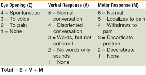
This scale is commonly used and was developed by Teasdale and Jennet. The Glasgow Coma Scale (GCS) was the first system to attempt to quantify the severity of a head injury.10 There are three variables used with the scale—best motor response, best verbal response, and eye opening. The key to calculating the GCS is to determine the best response in each category. The motor response is used to assess the level of central nervous system (CNS) function, whereas a verbal response shows the ability of the CNS to integrate information. Eye opening will demonstrate brainstem activity. Each category is given a score, as outlined in Table 4-1, with total scores ranging from 3 to 15. Higher scores represent increased levels of consciousness. The letter T is used to designate that the patient was intubated at the time of examination. A GCS of 8 or less is generally accepted as a definition of coma. At this level, there will often be a monitor placed to measure intracranial pressure (ICP) because it is difficult to monitor these patients for neurologic deterioration as compared with a patient with a higher GCS. Of patients with a moderate head injury (GCS, 9 to 13), approximately 10% to 20% will deteriorate and lapse into a coma. This can influence the timing of the management of maxillofacial injuries. Unless the maxillofacial injures compromise the airway, these patients are not routinely intubated as long as the airway can be protected. For patients with a severe head injury (GCS, 3 to 8), urgent management is critical. These patients are at the greatest risk for mortality and morbidity. They will generally have an endotracheal tube placed to protect their airway. Airway protection can be complicated in the head-injured patient because there are often concomitant maxillofacial injuries.

TABLE 4-1

Glasgow Coma Score

image

Trauma Score and Revised Trauma Score.

The Trauma Score was initially developed in 1981 by Champion et al. The purpose of the score was to assess the extent of injury to vital systems and the severity of injury quickly to aid with triage and the treatment of the trauma patient.11 In 1989, Champion et al revised this scoring system, which became the Revised Trauma Score.12 The Trauma Score and Revised Trauma Score quantify the physiologic status of the patient’s respiratory, cardiovascular, and neurologic systems. The revised trauma score has a coded value for three variables—respiratory rate, systolic blood pressure, and GCS. Each variable is coded from 0 to 4, with a total score ranging from 0 to 12. Lower scores represent an increased severity of injury.

Injury Severity Score.

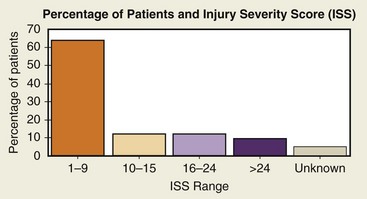
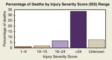
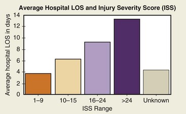
The Injury Severity Score (ISS) was developed to deal with multiple traumatic injuries to multiple organ systems. Scoring is based on the severity of injury to the three most injured organ systems, including respiratory cardiovascular systems, CNS, abdomen, extremities, and skin. Each of the three most injured organ systems is graded from 1 (minor) to 6 (fatal). The grades are then squared and added to come up with a total score from 3 (12 + 12 + 12) to 108 (62 + 62 + 62). The ISS has a practical range from 1 to 75; the risk of death increases with a higher score. ISS rates have been used to predict mortality rates by comparing ISS with mortality by age group. The National Trauma Data Bank (NTDB) uses this scoring system and categorizes an ISS from 1 to 9 as minor, 10 to 15 as moderate, 16 to 24 as severe, and more than 24 as very severe. Figures 4-4 and 4-5 show NTDB data as they relate to the ISS. The average length of stay increases approximately 3 days for each level of severity, as outlined in Figure 4-6.

Mechanism of Injury

The mechanism of injury can provide insight into other possible injuries that have not yet resulted in significant changes in vital signs or physiologic function. The mechanism of injury is usually one of the first issues communicated by EMS to the trauma team as a patient enters the trauma bay. Factors with a high correlation with life-threatening injuries are outlined in Box 4-1.

Other Factors

Various anatomic factors that correlate with high mortality include those listed in Box 4-2. Other factors that have been shown to worsen prognosis of a trauma patient with only a moderately severe injury include concurrent disease, age younger than 5 years or older than 55 years, and cardiac or respiratory disease.

Primary Survey

Once the trauma victim arrives at the resuscitation room, a quick assessment of major injuries, vital signs, and mechanism of injury is done to prioritize what needs to be treated first. The airway is always the first concern. A trauma team leader will command the personnel regarding actions to take. A team member will be ready to write all the important information regarding the patient who has just arrived. There are formats specially designed for this purpose with human body graphics in different views, whereby drawings could be made for better documentation of the injuries. EMS personnel usually will describe the information regarding the patient as they enter the room.

The treatment could be summarized as primary survey, resuscitation of vital functions, a second survey, once the patient has stabilized, and definitive care. The logical sequence of this process constitutes the ABCDEs of trauma:

During the primary survey, the life-threatening conditions are identified and treated and vital signs are stabilized. Different conditions are frequently treated simultaneously.

Special consideration must be given to pediatric, pregnant, and older patients. In general, the guidelines are the same, but the health care provider should take into consideration the differences in physiology, metabolism, response to trauma, amount of fluids, resuscitation techniques, and medication dosage. In the pregnant patient, the condition can be assessed by palpating the abdomen or a performing a human chorionic gonadotropin test. It is important to mention that in the geriatric population, comorbidities such as diabetes, cardiovascular disease, coagulopathies, liver and renal disease, and the long-term use of medications can alter the physiologic response to injury.

Airway Maintenance with Cervical Spine Control

On initial evaluation, ascertaining patency of the airway is of the highest priority. The perfusion of the brain and other vital structures will mean the difference between life and death, or permanent disability. Maintaining oxygenation and preventing hypercarbia are critical, especially for patients who have sustained head trauma. In a practical way, in a nonintubated patient, asking brief questions will lead to a quick evaluation of the airway and neurologic status. If the patient can talk, the airway is often patent. This assessment has to be repeated constantly.

The airway is especially critical in the head and/or maxillofacial trauma patient. The causes of upper airway compromise in the trauma patient may be tongue position, aspiration of foreign bodies, regurgitation of stomach contents, or facial, mandibular, tracheal and/or laryngeal fractures, bleeding, a retropharyngeal hematoma resulting from cervical spine fractures. or traumatic brain injury.13 Any laceration to the neck is classified according to the anatomic level and aggressively explored. Placement of a surgical airway may be necessary if endotracheal intubation is not possible.

A patent airway must be established while still protecting the cervical spine. A jaw thrust or chin lift procedure can be used to accomplish this. The jaw thrust maneuver involves the placement of the fingers of both hands on the angle of the mandible and the thumbs over the teeth or chin. The mandible is then gently pulled forward and rotated inferiorly. The elbow may be placed on the surface next to the patient to assist with stability. This procedure is the safest method of jaw manipulation in a patient with a suspected cervical spine injury. However, it does necessitate the use of both hands, and assistance is required to remove debris from the airway.

A chin lift procedure may be performed by placing the thumb over the incisal edge of the mandibular anterior teeth and wrapping the fingers tightly around the symphysis of the mandible. The chin is then gently lifted anteriorly and the mouth opened, if possible. Care should be taken not to extend the neck while the other hand should be used to clear any debris from the oral cavity.

Tonsillar suction should also be used to clear any secretions or other accumulations from the pharynx. A nasogastric tube or soft suction catheter may be used in patients without suspected substantial midface fractures or cranial base fractures because these tubes can be inadvertently passed into the cranial vault. An oral or nasal airway should be placed to keep the airway patent and the tongue elevated off the posterior pharynx. A nasal airway is better tolerated in an awake patient.

Any patient with multisystem trauma, altered level of consciousness, or blunt trauma above the clavicle should be assumed to have a cervical spine injury. Hyperextension or hyperflexion of the patient’s neck during airway establishment should be avoided. Excessive movement can turn a cervical spine injury without neuronal damage into neuronal deficit or even paralysis. In a multiple trauma patient with an altered level of consciousness, or any injury above the clavicle, the cervical spine (C-spine) has to be protected and stabilized. The presence of distracting injuries may deflect the attention to the C-spine; usually, a cervical collar is placed until the C-spine could be cleared. If during the placement of a definitive airway, the cervical collar has to be removed, another trauma team member will hold the head and neck to provide temporary stabilization. The cervical spine can be maintained in a neutral position using a backboard, bindings, and purpose built head immobilizers. The use of soft or semirigid collars is discouraged because they only stabilize 50% of movement. Cervical spine control should be maintained in a patient with suspected cervical spine injury until it can be ruled out clinically and/or radiographically during the secondary survey. Patients with severe head injury and an altered level of consciousness because of alcohol or drugs, or with a Glasgow Coma Score (GCS) of 8 or less, usually require the placement of a definitive airway with C-spine protection.

A patient who does not demonstrate purposeful motor responses or a patient who is combative often requires definitive airway management, with placement of an airway cuffed tube in the trachea and the tube connected to some form of oxygen ventilatory system. All airway management equipment should always be taped in place.

Endotracheal Intubation

When intubation is to be performed, two trained individuals should be involved, one performing the intubation and the other administering medications such as sedatives and muscle-paralyzing drugs, which should be used with caution. In many cases in which intubation is performed during the initial assessment, the use of sedatives and muscle blockers are unnecessary.

Oral or nasal intubation should be performed according to the operator’s preference and the injuries that are present. Extraordinary care must be taken in cases of facial trauma (e.g., nasal, frontal, midface fractures) In addition, applying cricoid pressure during the intubation will help to prevent aspiration.

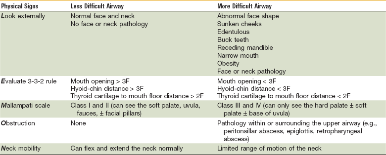
The mnemonic LEMON (Table 4-2) is useful when assessing the difficulty for intubation:

TABLE 4-2

LEMON Chart

image

After initial preoxygenation, a direct laryngoscopy is performed. The tube is inserted in the trachea and the cuff inflated; once the position of the tube is verified, it is secured and connected to a ventilation system. A gum elastic bougie can be used in cases of difficult intubation.

Airway control by rapid-sequence tracheal intubation (oral endotracheal tube [OETT]) is performed with in-line stabilization of the cervical spine. Correct placement of the endotracheal tube can be confirmed using the following:

The pediatric patient needs special consideration regarding instrumentation and knowledge of different anatomic features, such as the position of the larynx. In addition, obesity and retrognathia are other medical conditions for which intubation may be more challenging.

Several well-defined options for achieving airway control must be established in the event that OETT placement is not able to be achieved. These options include laryngeal mask airway (LMA), intubating LMA, fiberoptic intubation, percutaneous cricothyroidotomy, and surgical cricothyroidotomy (tracheostomy in children). Tracheal inspection is essential to determine whether there is peritracheal crepitus or deviation from the midline indicating potential direct airway, intrathoracic pulmonary, or major vascular injury.

Surgical Airway

When intubation is not possible, such as edema of the glottis, laryngeal fracture, or profuse hemorrhage, a surgical airway must be established. A cricothyrotomy is preferred to a tracheotomy because it involves less time and is associated with less bleeding.

Breathing

Once a patent airway is verified or established, pulmonary function should be assessed. Adequate exchange of gas is required for oxygenation and elimination of carbon dioxide. Gas exchange necessitates adequate ventilation. The lungs, chest wall, and diaphragm must all function adequately to ensure proper ventilation.

Breathing evaluation is most readily accomplished by visual inspection and palpation of thoracic cage movement and auscultation of gas entry. The patient is assessed for inequalities in chest movement from one side to the other, crepitus, and local movement asymmetry, as in paradoxic thoracic cage movement in flail chest. A trained provider should also be evaluating the patient for signs of impending respiratory failure, such as uncoordinated thoracic cage and abdominal wall movement, accessory muscle use, and stridor.

Inadequate ventilation may result in hypoxemia, hypercarbia, cyanosis, depressed level of consciousness, bradycardia, tachycardia, hypertension, and/or hypotension. As a general rule, until stability has been ensured, one should administer high-flow oxygen by mask to all patients to abrogate the potential for hypoxemia.

If the patient is breathing spontaneously and ventilation is adequate, supplemental oxygen can be administered by face mask. Assisted ventilation has to be instituted if opening of the airway does not result in spontaneous ventilation. Compromised ventilation could be the result of airway obstruction, altered mechanics, or CNS depression.

The exchange of air does not guarantee adequate ventilation. There are injuries that may impair ventilation, causing an obstructive or mechanical impairment. For example, a patient with a pneumothorax, flail chest, or hemothorax may have a chest wall that moves but ventilation may still be inadequate. In addition, shallow breaths or slow rates may not allow for adequate ventilation. Very slow and rapid rates of respiration suggest poor ventilation. Older patients with pulmonary dysfunction fall into a group with an increased risk of developing mechanical pulmonary problems.

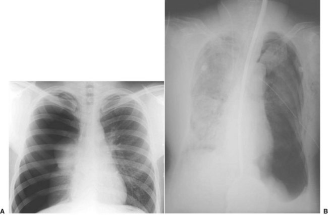
A patient’s respiratory status should be monitored constantly. Signs of ventilator deterioration warrant placement of a secured airway via an endotracheal tube or the initiation of assisted ventilation. At this point, a patient can be artificially ventilated by a bag-valve mask or a bag attached to an endotracheal tube. Patients who require assisted positive-pressure ventilation with an Ambu-Bag or mechanical ventilator should be closely monitored if their chest status has not been completely evaluated. A simple pneumothorax can be converted into a tension pneumothorax when the intrathoracic pressure increases (Fig. 4-7). In the presence of a pneumothorax, especially a tension pneumothorax, immediate treatment with a regular chest tube or needle insertion is necessary.

During the primary survey, the chest should be fully exposed and inspected for any signs of obvious injury. Presence of bruising, flail chest, penetration, and bleeding should be noted. The chest should be palpated for signs of rib or sternal fractures. Any subcutaneous emphysema should be appreciated. The neck should be evaluated for any sign of tracheal deviation and jugular venous distention. Chest expansion should be equal bilaterally, without intercostal or supraclavicular muscle retractions during respiration. The breathing rate should be assessed for signs of abnormality, such as tachypnea. Tachypnea with shallow respirations is suggestive of chest injury and impeding hypoxia. Distant heart sounds and distention of the neck veins can be suggestive of cardiac tamponade. If any of these conditions is suspected, before or after intubation and initial ventilation, a chest x-ray would be mandatory.

Head injuries may result in abnormal breathing patterns that could alter normal ventilation. Spinal cord injuries at the level of the cervical spine may affect normal muscle function and therefore compromise oxygen demands. Complete cervical transection at the C3 and C4 levels will compromise the phrenic nerves, resulting in abdominal breathing and paralysis of the intercostal muscles.

Standard monitors with a capnometer and pulse oximeter ensure adequate ventilation evaluation. Remember that the pulse oximeter does not measure the partial pressure of oxygen (PaO2) and, according to the position of the oxyhemoglobin dissociation curve, the PaO2 could vary. The use of pulse oximetry alone cannot distinguish between oxyhemoglobin and carboxyhemoglobin or methemoglobin. This is an important consideration in patients with vasoconstriction and carbon dioxide poisoning.

Circulation Management

In the primary survey, circulation becomes the priority after airway and breathing have been definitively managed. Delivery of oxygen to the tissues is dependent on adequate circulation. The main cause of deaths that can be prevented is caused by hemorrhage. It is estimated that hemorrhage accounts for 30% to 40% of trauma mortality, with 35% to 65% of deaths occurring in the prehospital period; 50% of deaths secondary to hemorrhage occur within the first 24 hours after the initial trauma.14 In general, blood volume is 7% of body weight; in children, it is considered to be 8% to 9%. Shock in a trauma patient is primarily hypovolemic secondary to trauma, although the patient may present with cardiogenic, neurogenic, or even septic shock. There are conditions that will contribute to a shocklike tension pneumothorax by reducing venous return. Extensive damage to the CNS or spinal cord may result in a neurogenic shock. In very unusual situations, septic shock may be present if the treatment of the patient was initiated several hours after the initial trauma.

Response to Shock

The initial circulatory response to hemorrhage is physiologic compensation. There is a release of endogenous catecholamines and hormones. The catecholamines will increase vascular resistance, increasing diastolic blood pressure and reducing pulse pressure—difference between the maximum and minimum blood pressures produced during one heartbeat.

Other vasoactive hormones that are released include histamine, bradykinin, beta endorphins, and a cascade of postanoids and cytokines. Vasoconstriction of the skin and extremities help maintain perfusion to the vital organs of the brain, kidneys, and heart. There will be an increase in heart rate to maintain cardiac output. Initially, blood pressure is not affected, although once compensatory mechanisms are overcome, it will be affected.

In cases of major fractures of the tibia or humerus, blood loss could reach up to 1.5 units. In femoral fractures, it could be twice that amount and, in pelvic fractures, blood loss could be significant, presenting as a retroperitoneal hematoma. Edema, in the case of increased endothelium permeability fluid loss, will shift from the plasma to the extravascular space.

Poor perfusion will affect aerobic metabolism, with nutrients and oxygen being deprived, and consequently result in anaerobic metabolism, with the formation of lactic acid. This will ultimately lead to metabolic acidosis. If this situation continues, the cell membrane will lose its integrity and there will be progressive cellular damage. Continuous hypoxia will result in cellular death. Research has shown that the lethal triad of acidosis, hypothermia, and coagulopathy initially cause the first problems in the polytrauma patient, associated with high mortality.15

The initial damage to soft tissues and organs, and fractures in the trauma patient, represents a major challenge. The local tissue damage resulting from contusions or lacerations, hypoxia, and hypotension result in further damage from local and systemic responses. These processes are activated to preserve immune system integrity and stimulate reparative mechanisms. This systemic inflammation is known as SIRS (systemic inflammatory response syndrome). In addition, the initial inflammatory response is augmented by a second hit, such as ischemia and reperfusion injuries and surgical interventions or infections (two-hit theory).16

In this chapter, the differences between adults and children are discussed, followed by a review of circulatory compromise and its definitive management. Causes of circulatory compromise include the following:

Effects of circulatory compromise include the following:

Assessment

The surgeon should be aware of the simultaneous compensatory mechanisms in a trauma patient and also the individual situation. In the older patient, for example, aging will affect general and specific organ systems that are predictive of failing health. Medically compromising conditions and the use of different medications are frequently present in this group and can contribute to high morbidity and mortality rates with relative minor injuries.17 According to the ABCs of trauma, once the airway is patent and ventilation is reestablished, the hemodynamic status (blood volume and cardiac output) should be assessed, as follows

Rapid pulse may indicate blood loss whereas an irregular pulse may indicate cardiac dysfunction. Different age categories should be taken into consideration when assessing heart rates. For example, young patients with normal vagal tone and older patients with pacemakers and/or beta blockers will have different responses to hypovolemia. The heart rate will not increase as expected.

• Respiratory rate: According to the degree of hemorrhage present, patients may become tachypneic as a physiologic response to the need for more oxygen to be delivered to the tissues.

• Blood pressure: If the blood loss is significant (>30% of blood volume), there will be changes in blood pressure.

• Skin color: A gray, pale ashen tone may indicate hypovolemia; pink skin is an indication of good perfusion.

• Urinary output: Urinary output is considered to be in normal limits with approximately 0.5 mL/kg/hr for the adult and 1 mL/kg/hr for children. A decrease of urinary output to less than 30 mL/hr in an adult may indicate hypovolemia in the absence of other medical conditions (e.g., renal damage).

•&nb/>

Stay updated, free dental videos. Join our Telegram channel

Jan 14, 2015 | Posted by in Oral and Maxillofacial Surgery | Comments Off on 4: Initial Assessment and Intensive Care of the Trauma Patient

VIDEdental - Online dental courses

Get VIDEdental app for watching clinical videos