32
Impacted maxillary canines
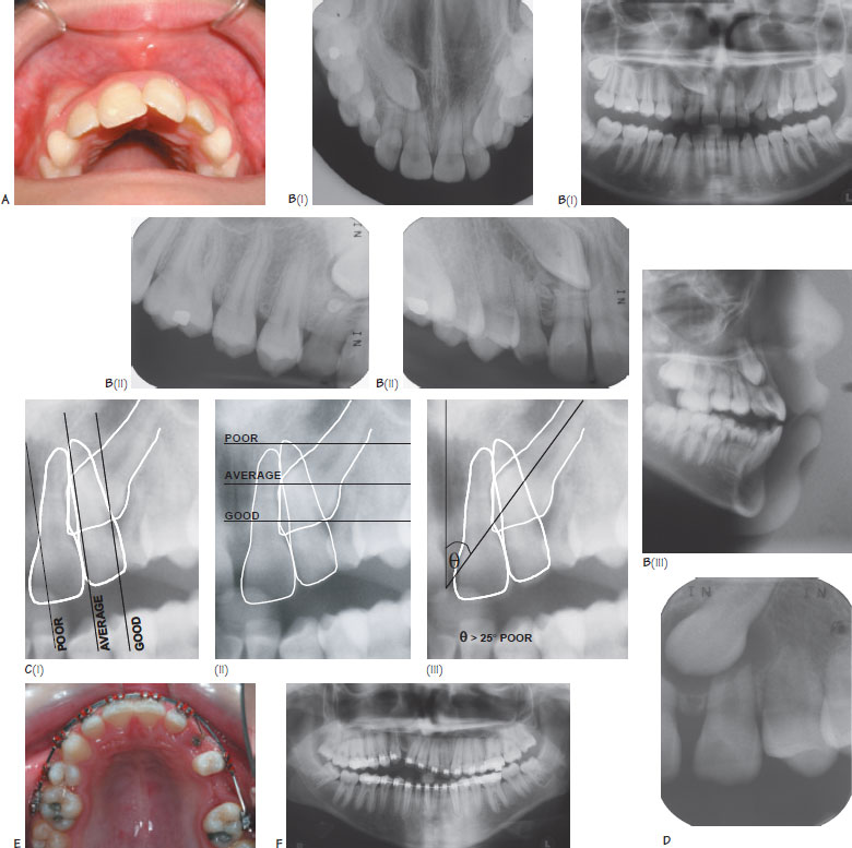
Figure 32.1 (A) The canines should be palpable buccally by the age of 10 years. Note the canine bulge in the figure. If the canines are not palpable, one should consider radiographic examination to determine their position. (B) Radiographic localisation of impacted maxillary canines, (i) Vertical parallax using a DPT and upper anterior occlusal radiograph. Note that the canine moves in the direction of the tube shift indicating a palatal position, (ii) Horizontal parallax using two periapical views taken at different angles. Note the canine moves in the direction of the tube shift, (iii) A lateral cephalogram can also be used to determine the anteroposterior and vertical position of an impacted canine. (C) Assessment of the prognosis for canine alignment, (i) The anteroposterior (AP) position can be assessed by drawing a line mesial to the central and lateral incisor and distal to the lateral incisor. The position of the canine crown in relation to these lines indicates the prognosis for alignment in the AP plane, (ii) The vertical position can be assessed by dividing the lateral incisor root into thirds. The position of the canine crown in relation to these lines indicates the prognosis for alignment in the vertical plane, (iii) The angulation of the long axis of the canine can be measured in relationship to a vertical line. An angulation of >25° indicates likely difficulty with alignment. (D) The incisor roots are at risk of resorption in the presence of palataIly ectopic canines. In this example, both the central and lateral incisor roots were resorbed. (E) Alignment of an impacted maxillary left canine using a fixed appliance. The canine has been exposed and bonded and an archwire is being used to apply orthodontic traction. (F) The upper right maxillary canine has become ankylosed and has failed to align following the application of orthodontic forces. Note how the adjacent teeth have intruded in response to the orthodontic forces.
Following the third molars, the maxillary canine is the most commonly impacted tooth. The prevalence of maxillary canine impaction is 1–2% with a female:male ratio of 2:1. Eighty-five per cent of impactions are palatal whilst the remainder (15%) occur buccally.
Normal development of the maxillary canine
The maxillary canine begins its development high in the maxilla at the age of 4–5 months. The tooth has a long path of eruption, passing along the distal root surface of the lateral incisor, buccal to the deciduous canine to its final position with eruption at 11–12 years. The canine should be palpable high in the buccal sulcus by the age of 10 years (Figure 32.1A).
Stay updated, free dental videos. Join our Telegram channel
VIDEdental - Online dental courses


