3: Immunity and Immunologic Oral Lesions

Immunity and Immunologic Oral Lesions

Margaret J. Fehrenbach, Joan Andersen Phelan and Olga A.C. Ibsen

Objectives

After studying this chapter, the student will be able to:

1. Define and pronounce each of the words in the vocabulary list for this chapter.

2. Describe the differences between an immune response and an inflammatory response.

3. List and describe the two main types of lymphocytes, including their origins and activities.

4. List and describe the different types of T-cell lymphocytes and their functions.

5. Describe the origin of macrophages and list their activities in the immune response.

6. Describe the differences between humoral immunity and cell-mediated immunity and include the cells involved in each.

7. Describe the differences between passive and active immunity and give an example for each type of immunity.

8. List and describe four types of hypersensitivity reactions and give an example for each type of hypersensitivity.

9. Describe the process of autoimmunity and how it results in disease.

10. Describe the process of immunodeficiency and how it results in disease.

11. Describe and contrast the clinical features of each of the three types of aphthous ulcers.

12. List systemic diseases associated with aphthous ulcers.

13. Describe and compare the clinical features of urticaria, angioedema, contact mucositis, and fixed drug eruption.

14. Describe the clinical features of erythema multiforme and Stevens-Johnson syndrome.

15. Describe the clinical and microscopic features of lichen planus.

16. List the triad of systemic signs that comprise reactive arthritis (Reiter syndrome) and describe the oral lesions that occur in this condition.

17. Name the two cells that characterize Langerhans cell disease microscopically. Describe the acute disseminated form, chronic disseminated form, and chronic localized form and state the names that traditionally have been used for each of these conditions.

18. Describe the oral manifestations of each of the following autoimmune diseases: lupus erythematosus, pemphigus vulgaris, mucous membrane pemphigoid, and Behçet syndrome.

19. Define desquamative gingivitis, describe the clinical features, and list three diseases in which desquamative gingivitis may occur.

20. Describe the clinical features of Behçet syndrome.

Vocabulary

The inflammatory response, as described in Chapter 2, is a rapid first line of defense against injury. The immune response, described in this chapter, follows the inflammatory response and is necessary for complete recovery. This chapter begins with a description of the acquired immune response and then includes a discussion of the most commonly encountered oral diseases that may result from harmful effects of such a response. Surveillance against neoplastic cells also involves the immune response and is described, along with the neoplastic process and neoplastic oral lesions, in Chapter 7.

Acquired Immune Response

Like the inflammatory response, the acquired immune response defends the body against injury, particularly from foreign substances such as microorganisms. The immune response differs from the inflammatory response in that it has the capacity for memory and responds more quickly to a foreign substance if encountered again. It works amid the background of an already activated inflammatory response and innate immune response, as well as a working repair process. The innate immune response does not involve memory and is described, along with inflammation, in Chapter 2. In contrast, the acquired immune response involves a complex network of white blood cells. Similar to the inflammatory response, the immune response may also result in an increased level of tissue damage and disease as it fights against what it considers foreign.

Antigens

Antigens, or immunogens, are foreign substances against which the immune system defends the body. These substances are mainly proteins and are often microorganisms and their toxins. The immune system tolerates the normal components of the body, or self, with their normal diversity. In contrast, antigens are substances that the immune system recognizes as foreign, or nonself. Transformed human cells such as neoplasm cells or cells infected with viruses can become antigens. Human tissue, as in the case of an organ transplant, tissue graft, or incompatible blood transfusion, can also become an antigen.

In one type of disease, an autoimmune disease, parts of an individual’s own body become antigens. In another type of disease, an immunodeficiency, the body no longer recognizes certain antigens as foreign. In another type of reaction involving hypersensitivity, the body overreacts to what it sees as foreign, creating a multitude of complications. Thus disease can occur when the immune response identifies components of self as antigen, does not recognize foreign material as antigen, or overreacts to antigens.

Cellular Involvement in the Immune Response
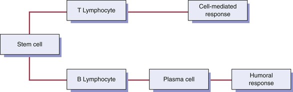
A complex network of white blood cells is involved in the immune response. These cells can produce cell products, or cytokines, that are active during the immune response. The primary white blood cells involved in the immune response are lymphocytes. These cells are able to recognize and respond to an antigen when it is in contact with receptor sites located on the cell surface. Lymphocytes, like other white blood cells, are derived from stem cells in the bone marrow. Lymphocytes constitute 20 to 25% of the white blood cell population. They are mobile antigen-sensitive cells with a long life. They have a round nucleus and only a small amount of cytoplasm. Different types of lymphocytes have differing functions. There are three main types of lymphocytes: (1) the B-cell lymphocyte, (2) the T-cell lymphocyte, and (3) the natural killer cell. The macrophage and the dendritic cell are also part of the immune response.

B-Cell Lymphocyte

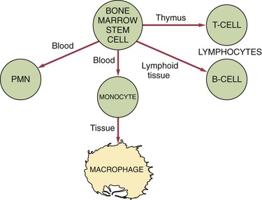
The B-cell lymphocyte (B cell) develops from a stem cell in the bone marrow and then resides and matures in lymphoid tissue (Figure 3-1). When antigen stimulates a B cell, the B cell travels to the site of the injury. Two main types of B cells develop when stimulated by an antigen: (1) the B-memory cell and (2) the plasma cell.

The B-memory cell retains a memory of the antigen. In the presence of an antigen recognized by the B-memory cell, this cell reacts by duplicating itself many times over in a process called clonal selection. All of these newly formed B cells retain the capacity to recognize the previously encountered antigen.

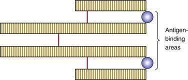
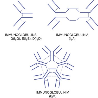
The plasma cell has a round, pinwheel-shaped nucleus and visible cytoplasm. The plasma cell produces and releases many copies of a particular protein (antibody) in response to the presence of antigen. Antibodies circulating within the blood serum are considered immunoglobulins. Five different general types of immunoglobulins exist: (1) IgA, (2) IgD, (3) IgE, (4) IgG, and (5) IgM (Table 3-1). They all are variations of the same basic structure (Figures 3-2 and 3-3). Each immune response involves specific antibodies produced in response to a specific antigen by a specific plasma cell. This specificity is a characteristic function of the immune response. The level of a specific antibody in the blood is called the antibody titer. This can be measured by diagnostic laboratory tests and is useful in the diagnosis and evaluation of infectious disease.

TABLE 3-1

Immunoglobulins (Antibodies)

IMMUNOGLOBULIN (IG) DESCRIPTION
IgA Has two subgroups: serous in the blood and secretory in the saliva and other secretions such as tears and breast milk; aids in defense against proliferation of microorganisms in body fluids as well as protecting mucosal sites (e.g., gastrointestinal and genitourinary tracts)
IgD Functions in the activation of B-cell lymphocytes, because it is found on their surface, but full role is unclear
IgE Involved in hypersensitivity or allergic reactions because it can bind to mast cells and basophils to bring about the release of biochemical mediators such as histamine; and also attacks parasites
IgG Major antibody in blood serum (about 75%); can pass the placental barrier, produced in secondary immune response, and serves as the first passive immunity for the newborn
IgM Involved in early immune responses because of its involvement with IgD in the activation of B-cell lymphocytes, activates complement, and reacts to blood group antigens

The combination of an specific antibody with a specific antigen is an immune complex. The formation of an immune complex usually renders the antigen inactive. Immune complexes may also be involved in certain disease states. Some of these are discussed later in this chapter.

T-Cell Lymphocyte

After it develops from a bone marrow stem cell, the T-cell lymphocyte (T cell) travels to the thymus and is processed into a mature cell (see Figure 3-1). The thymus is a primary lymphoid organ located high in the chest. It is quite large in infants and shrinks as an individual matures.

The T cell can be distinguished from other lymphocyte types, such as B cells and natural killer cells, by the presence of a special receptor on its cell surface that is called the T-cell receptor (TCR). Other receptors are also present on the surface of the T cell. Different types of T cells have different functions in the immune response. The different types of T cells include the T-helper cell, the T-suppressor cell, the T-cytotoxic cell, and the T-memory cell.

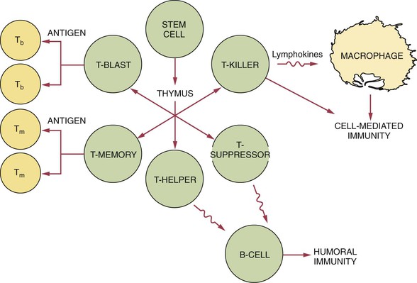
The T-helper cell increases the functioning of the B-cell, enhancing the antibody response. It is easily identifiable by the CD4 cell receptor on its surface (Figure 3-4). The T-suppressor cell (or T-regulator cell) carries the CD8 marker as well as other markers on its surface and suppresses the functioning of the B cell.

The T-cytotoxic cell also carries the CD8 marker. It is active in surveillance against virally infected cells and neoplastic cells, directly attacking these cells.

T cells within cell-mediated immunity also start, regulate, and coordinate the overall immune response.

Macrophage

Not only is the white blood cell macrophage present in the connective tissue during inflammation, it is also involved in the evolving immune response to an antigen as an accessory cell (see Figure 3-1). The macrophage is active in phagocytosis of foreign substances and also assists both the B-cell and T-cell during the immune response.

Along with phagocytosis, the macrophage acts to process and present antigen material on its surface to the T-helper cell. This stimulates both types of lymphocytes to travel from the lymphoid tissue or surrounding blood vessels to the injury site. Thus the macrophage functions as an antigen-presenting cell, acting as a messenger between both the inflammatory response and immune response.

The macrophage carries receptors for lymphokines produced by the lymphocytes, which allow it to be activated into a single-minded pursuit of specific foreign material such as microorganisms or neoplastic cells (Table 3-2). When activated, the macrophage can function in many other different ways, always amplifying the immune response. Unlike lymphocytes, the macrophage does not retain memory of an encountered antigen and needs to be reactivated during each encounter.

TABLE 3-2

Cytokines

CYTOKINE FUNCTION
Interferons Various functions involving leukocytes, fibroblasts, and endothelial cells
Interleukins Stimulate leukocyte proliferation and other functions
Lymphotoxin Destroys fibroblasts
Macrophage-activating factor Activates macrophages to produce and secrete lysosomal enzymes
Macrophage chemotactic factor Stimulates macrophage emigration
Migration inhibitory factor Inhibits macrophage activity
Tumor necrosis factor Various functions involving leukocytes and fibroblasts

Dendritic Cell

The dendritic cell (DC) is a white blood cell whose main function is to process antigenic material and present it on its surface to other cells of the immune system. Thus the dendritic cell functions as an antigen-presenting cell, acting as a messenger between innate immunity and acquired immunity. It is similar in function to a macrophage.

The dendritic cell is present in tissue that is in contact with the external environment, such as the skin and mucosa. It can also be found in an immature state in the blood. At certain developmental stages, it grows branched projections or dendrites. Once activated, it migrates to the lymph nodes or other lymphoid tissue, where it interacts with T cells and B cells to initiate and shape the acquired immune response. In mucosal tissue, including the oral mucosa, there is a specialized dendritic cell type called a Langerhans cell.

Cytokines

Cytokines are proteins that are made by cells and are able to affect the behavior of other cells; thus they are considered immunomodulating agents. Immunomodulating agents, or immunomodulators, alter the immune response by augmenting or reducing the functions of the response. Cytokines are produced by the cells of the immune system and play a prominent role in the activation of the immune response. They are one way that lymphocytes communicate with each other and with other immune system cells. B cells and T cells produce their own cytokines, called lymphokines, in varying levels (see Figure 3-4).

Different cytokines have differing functions within the immune response (see Table 3-2). They can activate macrophages and enhance the ability of macrophages to destroy foreign cells. These types of cytokines may also be involved in various other functions concerning leukocytes, fibroblasts, and endothelial cells. They are also responsible for the systemic effects of inflammation such as loss of appetite and increased heart rate. Monocytes/macrophages produce their own cytokines, called monokines. Dendritic cells, such as the Langerhans cells, also produce and react to cytokines.

One of the first cytokines to be discovered within the body was interferon. Produced by T cells and macrophages (as well as by cells outside the immune system), interferons are a group of proteins with antiviral properties. Chemokines are a group of cytokines that are named for their ability to induce chemotaxis in nearby responsive cells. They may be involved in controlling infection as part of the immune response as well as being involved in several developmental processes during embryogenesis.

Major Divisions of the Immune Response

There are two major divisions of the immune response: (1) humoral immunity and (2) cell-mediated immunity (Figure 3-5). These two divisions differ in their reaction to an antigen. However, these divisions are interrelated, and present understanding of the immune response recognizes that these divisions do not function as distinctly separate mechanisms.

Humoral immunity, or antibody-mediated immunity, involves the production of antibodies, with the B-cell lymphocytes as the primary cells. Humoral immunity is responsible for protection against many pathogenic microorganisms such as bacteria and viruses.

The other division of the immune system is cell-mediated immunity, or cellular immunity; it involves lymphocytes, usually T cells, working alone or assisted by macrophages. The cell-mediated division regulates both major divisions of the immune system.

Types of Immunity

Two types of immunity can occur: (1) passive and (2) active. Passive immunity refers to the use of antibodies produced by another person to protect an individual against infectious disease. This type of immunity can occur naturally or it can be acquired. Natural passive immunity occurs when antibodies from a mother pass through the placenta to the developing fetus. These antibodies protect a newborn infant from disease while the infant’s own immune system matures.

Passive immunity can also be acquired by injecting a person with antibodies against a microorganism to which the person has not previously been exposed. This is done to confer immediate protection against the disease caused by that microorganism. These antibodies are collected from individuals who have already had the disease and have naturally produced antibodies to the pathogenic microorganism. Acquired passive immunity is short-lived but can act immediately. It is used because the unprepared immune system of an individual takes longer to produce antibodies, and in the meantime the disease may develop. Acquired passive immunity may be provided to dental personnel who do not have immunity to hepatitis B after needlestick or other occupational exposure incidents, using hepatitis B immunoglobulin.

Active immunity uses antibodies produced by one’s own body to protect against infectious disease, and it can occur naturally or be acquired. It occurs naturally when a pathogenic microorganism causes disease. Protection against further attack by that microorganism is conferred to the individual if the body recovers from the disease. A less risky way of achieving active immunity is by an acquired or artificial means. This production of acquired active immunity is called immunization. A person is injected with or ingests either altered pathogenic microorganisms or products of those microorganisms. The altered microorganism or products of the microorganisms cannot produce infection but is able to act as an antigen. This is called a vaccine, and the process is called vaccination. When the pathogenic microorganism is encountered after vaccination, the immune system produces a stronger, faster response and prevents development of the disease.

Immunization lowers the risk of a microorganism causing disease because it safely prepares the immune system to fight future attacks by the disease-causing microorganism. In some cases more than one exposure to the antigen is needed to ensure adequate immunity; a repeated exposure by way of a vaccination is called a booster. Immunization by vaccination is used to protect children and adults against many diseases. Dental personnel should be vaccinated against the hepatitis B virus because of their high risk of occupational exposure to that virus.

Killed-type vaccines consist of heat- or chemical-treated microorganisms or toxins; these treatments make killed-type vaccines safe but in some cases they may be less effective than live-attenuated vaccines. Live-attenuated vaccines consist of genetically altered microorganisms that have lost virulence but still undergo limited replication. Molecular vaccines are composed of critical antigenic determinants, derived as recombinant (from cloned bacteria or yeast) or synthetic peptides; adjuvants may be used to stimulate immune responses.

Immunopathology

The immune response helps defend the body against disease-producing antigens, but it can also malfunction and cause tissue damage resulting in the production of lesions. Immunopathology is the study of diseases caused by the malfunctioning of the immune system. These immunopathologic conditions include (1) hypersensitivity, (2) autoimmune disease, and (3) immunodeficiency. Examples of each of these immunopathological conditions, in which damage is directly caused by the immune response, are discussed next. Some of these conditions fall into the category of a syndrome, which is a group of signs and symptoms that occur together to define the disease state. Many different syndromes are described in various chapters in this text.

Hypersensitivity

Hypersensitivity or allergy comprises the same basic types of reactions that occur when the immune response is fighting microorganisms and protecting the body against disease. However, these hypersensitivity or allergenic reactions are exaggerated immune responses causing an immunopathologic condition, along with tissue destruction. Four main types of hypersensitivity reactions occur; they are classified by the nature of the immune response that causes the disease (Table 3-3).

TABLE 3-3

Hypersensitivity Reactions

TYPE OF REACTION EXAMPLE(S)
Type I or anaphylactic type Hay fever, asthma, anaphylaxis
Type II or cytotoxic type Autoimmune hemolytic anemia
Type III or immune complex type Autoimmune diseases such as systemic lupus erythematosus
Type IV or cell-mediated type Granulomatous diseases such as tuberculosis as well as graft and organ transplant rejection

Type I Hypersensitivity

Type I hypersensitivity or anaphylactic type hypersensitivity is a reaction that occurs immediately—within minutes—after exposure to a previously encountered antigen, or allergen in this case, such as pollen, latex, or penicillin. With type I hypersensitivity plasma cells produce IgE as a response to the allergen. The newly produced IgE binds to mast cells located in tissue, causing them to release their granules containing histamine, a potent biochemical mediator of inflammation. This results in edema caused by increased dilation and permeability of blood vessels and in constriction of smooth muscle in the bronchioles of the lungs.

This type of hypersensitivity includes hay fever, urticaria (hives), and more serious conditions, including asthma and anaphylaxis. Anaphylaxis is a type of hypersensitivity that can be life-threatening because the individual may not be able to breathe as a result of the oropharyngeal tissue swelling and constriction of the bronchioles, which requires immediate treatment with epinephrine.

Drugs that suppress inflammation, such as antihistamines and corticosteroids, may be helpful in reducing symptoms of some forms of type I hypersensitivity.

Type IV Hypersensitivity

Type IV hypersensitivity or cell-mediated type hypersensitivity involves a cell-mediated immune response rather than a humoral response that produces antibodies. T-cell lymphocytes that have been introduced to an antigen previously either directly cause damage to the tissue cells or recruit other cells that cause the damage. This type of hypersensitivity reaction is also called delayed hypersensitivity and is used when diagnosing tuberculosis. A visible skin reaction occurs if the individual tested has previously been exposed to the microorganism that causes tuberculosis.

This type of hypersensitivity is also responsible for the rejection of tissue grafts and transplanted organs as well as an allergy to nickel in dental restorations. New strategies for prevention of rejection, such as synthetic production of therapeutic antibodies against specific T-cell receptors, may produce fewer long-term side effects than the chemotherapies now routinely used.

Hypersensitivity to Drugs

Drugs can act as allergens and cause a hypersensitivity reaction. Many factors influence the risk of a hypersensitivity or allergic reaction to a drug. The route of administration influences how the reaction will be manifested and its severity. Topical administration of drugs (via the skin or mucous membranes) may cause a greater number of reactions than the oral (swallowed) or parenteral (administered by injection) route of administration; however, the reaction that occurs after parenteral administration may be more widespread and severe because the allergen can be carried quickly to many parts of the body by the circulating blood.

Patients with multiple allergies are more likely to have allergic reactions to drugs, and patients with autoimmune diseases such as systemic lupus erythematosus commonly have adverse reactions to medication. Children, with their newer levels of immunity, are less likely than adults to have an allergic reaction to a drug.

Drugs can be involved in any of the previously described hypersensitivity reactions. Type I hypersensitivity to a drug can include anaphylaxis, urticaria (hives), and angioedema (localized swelling). A systemic anaphylactic reaction is more likely to occur with an injected drug but can also occur with a drug administered orally, and can be fatal. For example, the drug penicillin may cause a systemic anaphylactic reaction in approximately 1 in 10,000 patients and causes about 300 deaths per year in the United States.

The classic example of type III hypersensitivity is serum sickness, which involves a drug allergy. This name was given to a reaction that occurred frequently when patients were given large amounts of horse antitoxin serum to provide passive immunity in the treatment of diphtheria and tetanus. This is no longer the method by which passive immunity to these diseases is provided. The drug penicillin is the single most common cause of serum sickness. Other drugs such as barbiturates can also cause this reaction. The signs and symptoms of serum sickness include rash or urticaria, fever, painful swelling of the joints (arthritis), renal disturbance or failure, edema around the eyes, and cardiac inflammation. Drugs can also be involved in a type IV hypersensitivity reaction. This T-cell–mediated allergic reaction can occur in response to topically applied substances and can produce contact dermatitis (skin inflammation) and contact mucositis, which is an inflammation of the mucosal tissues.

Autoimmune Diseases

The immune system learns to differentiate between one’s own cells or tissue and foreign substances early in embryologic development. This recognition and the nonresponsiveness of the immune system to one’s own cells or tissue usually produce a type of immunologic tolerance.

In an autoimmune disease the recognition mechanism breaks down, and certain body cells are no longer tolerated. The immune system now treats body cells as antigens, creating an immunopathologic condition. An autoimmune disease may involve a single cell type or a single organ or may be even more extensive, involving multiple organs. Certain types of tissue and even entire organs may be damaged. Genetic factors may play a role in the predisposition of an individual to autoimmune disease, and viral infection may also be involved. Some autoimmune diseases are also called connective tissue diseases. Several autoimmune diseases have oral manifestations and are described later in this chapter.

Oral Immunologic Lesions and Diseases

Aphthous Ulcers, Recurrent Aphthous Ulcers

Recurrent aphthous ulcers, also known as canker sores or aphthous stomatitis, are a painful type of oral ulcers for which the cause remains unclear. Aphthous ulcers are one of the most common oral lesions, occurring in about 20% of the general population. They frequently occur in episodes. The first episode of these ulcers usually occurs in adolescence, and they are somewhat more common in females than in males. The clinical appearance and location are important in establishing the diagnosis.

Trauma is the most commonly reported precipitating factor in the development of aphthous ulcers. They are often reported to occur after trauma to the oral mucosa during dental procedures (e.g., in the area of film or sensor placement or at the injection site for local anesthetics) or with the manipulation of oral tissue during dental hygiene treatment. Some patients associate the initiation of aphthous ulcers with eating certain foods such as citrus fruits. However, it is possible that patients perceive these foods as causative because of the sensitivity of the ulcers to foods with a high acid content, or the foods themselves may have caused trauma to the oral mucosa. The recurrence of aphthous ulcers has been associated with menstruation, whereas pregnancy has been found to produce a decrease in the episodes. These ulcers also occur in association with certain systemic diseases as well as with tobacco cessation. Emotional stress has also been suggested as a contributing factor.

Substantial evidence indicates that aphthous ulcers have an immunologic pathogenesis. Patients in whom aphthous ulcers develop have slightly elevated levels of antibodies to oral mucous membranes. Microscopically, an infiltrate of lymphocytes is present in the lesion, suggesting that cell-mediated immunity may be important in development of the ulcers. The infiltrate contains mainly T-helper cells in the prodromal stage and T-cytotoxic cells in the ulcerative phase; T-helper cells return in the healing s/>

Stay updated, free dental videos. Join our Telegram channel

Jan 12, 2015 | Posted by in Oral and Maxillofacial Pathology | Comments Off on 3: Immunity and Immunologic Oral Lesions

VIDEdental - Online dental courses

Get VIDEdental app for watching clinical videos