14 Pigmented lesions: Ethnic pigmentation and tattoos
Figure 14.1 Gingival racial pigmentation (very mild).
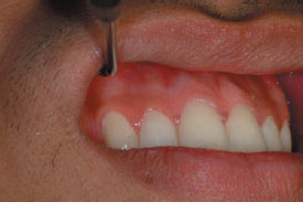
Figure 14.2 Buccal racial melanosis.
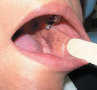
Figure 14.3 Amalgam tattoo.
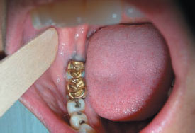
Figure 14.4 Foreign body tattoo following an explosion.
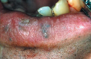
Figure 14.5 Submucosal amalgam may cause amalgam tattoo (patient from Figure 14.3).
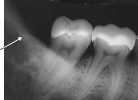
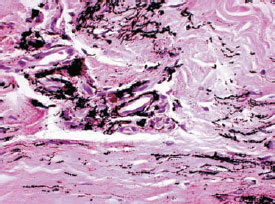
Figure 14.6 Amalgam tattoo × 40.
Ethnic pigmentation
Definition: Physiologic oral pigmentation manifests as multifocal or diffuse melanin pigmentation seen mainly in various ethnic groups, especially people of African or Asian heritage, but can also be noted in patients of Mediterranean descent, sometimes even in quite lightskinned people.
Prevalence (approximate): Occurs in all races. The intensity and distribution of racial pigmentation is variable, not only between races, but also between different individuals of the same race and within different areas of the same mouth.
Age mainly affected: All ages.
Gender mainly affected: M = F.
Etiopathogenesis: The darker a person’s skin color the more likely they are to have oral pigmentation. Skin color is a quantitative polygenic trait that varies continuously on a gradient from dark to light. Genes known to contribute to skin color are the MC1R and SLC24A5 genes.
Diagnostic features
History: May be first noticed in adult life and then assumed incorrectly to be acquired rather than congenital.
Clinical features: Brown or blackish patches most obvious in the anterior labial gingivae (Figu/>
Stay updated, free dental videos. Join our Telegram channel
VIDEdental - Online dental courses


