Chapter 10
The surgical microscope
Introduction
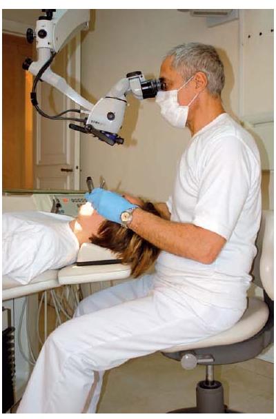
The desire to see better in order to address the complexity of the root canal system of teeth by endodontics has naturally led to the use of optical aids. Simple magnifying glasses (conventional Galilean optics) or magnifying glasses mounted on spectacles make it possible to achieve magnification up to 5×. Beyond these enlargements, optical systems become too heavy and cumbersome for them to be used for long periods without discomfort. If a source of light is fitted, the weight of the device increases even further. Although these measures for magnification are distinct improvements beyond the naked eye, the surgical microscope offers a stereoscopic, three- dimensional, enlarged image under bright illumination at a comfortable working position that will greatly enhance the precision of endodontics (Fig. 10.1). Thereby root canal orifices can be more easily found (Figs 10.2 and 10.3a–c), cracks and fractures revealed (Fig. 10.4), and instrumentation and filling procedures in both conventional (Fig. 10.3d–f) and surgical endodontics (Fig. 10.5) facilitated. This short chapter describes the principal uses and applications of the surgical microscope in endodontic therapies.
Components
The optical system
The surgical microscope intended for dental operations consists of three key components: an ocular head (viewing tube) holding two eyepieces, intermediate magnification lenses and the main objective lens (Fig. 10.6).
The focal length of the eyepieces lies between 100 mm and 160 mm, with magnification values from 10× to 12.5×. The eyepieces comprise an adjustment ring with settings to compensate for visual defects. The distance between the eyepieces is adjustable in order to match the appropriate interpupillary distance of the eyes. To be functional in most clinical situations it should be possible to tilt the ocular head in the frontal plane.
The magnification lenses placed between the eyepieces and the main objective make it possible to have several magnifications (generally from 5× to 25×). They are optical blocks mounted on a turret, from which, with the aid of a ring, the magnification most appropriate to the sequence of the operation can be selected. On the most sophisticated models this variation is controlled via a zoom lens.
The lens nearest to the object to be examined, the main objective lens, may have a varying focal length, chosen depending on the height and the working habits of the operator. It can vary from 200 mm to 300 mm and determines the working distance (distance from object observed to the objective lens). See Advanced concept 10.1.
The mechanical system
A suspension system is provided to ensure stability and balance inherent movements of the microscope away from the position in which it is originally placed, otherwise major operating difficulties would ensue. The system of arms is either mounted on a movable floor stand or fixed either to the wall or to the ceiling of the surgery. On purchasing an operating microscope the flexibility and ability of this mechanical system to offset movements is a determining factor. Where the working conditions allow, a ceiling mount is the preferred choice.
Fig. 10.1 Working position with the surgical microscope.
The illumination system
The light system consists of either a 150 watt halogen lamp or a xenon arc lamp, which gives comfortable cold, white light. The light is conducted by fiber optics and genera/>
Stay updated, free dental videos. Join our Telegram channel
VIDEdental - Online dental courses


