Figure 3.19 Slight facial asymmetry with prominence of chin deviated to patient’s left. There was no functional or symptom history significance to this asymmetry, and the asymmetry was judged to be within normal range.
B. Symptom History
- Thirty-five years prior, she was the passenger in a car involved in a motor vehicle collision; her car was hit from behind at the same time as her head was turned to the side window and she had a whiplash trauma. Within a month, she began to experience facial pain and mechanical problems of the jaw and was no longer able to open as wide. Functionally, she could no longer bite into a whole apple.
- Three years later the right TMJ received a silastic implant and the left TMJ received a disc repair. New symptoms emerged in the right TMJ, and the silastic implant was removed; residual scar tissue was allowed to replace the disc.
- Following implant removal, chewing triggered minor pain and some limitation, but otherwise her jaw condition was self-managed well.
- About 2 months prior to this consultation, a fractured mandibular tooth underlying a loose crown was extracted as part of a long dental appointment, and postoperative jaw opening was limited to a few millimeters; this gradually improved over about 6 weeks, during which her symptoms were affected by chewing, opening, and other jaw activities. Now her jaw opening has returned to her “normal.”
C. Medical History
- Occipital headaches, attributed to cervical spine arthritis. Previous 10-year history of bad headaches following the motor vehicle collision 35 years ago.
- Review of systems for eyes, ears, sinus, and teeth is negative.
- Cervical mobility is without complaint, though left upper extremity paresthesias occur on occasion, attributed to the cervical arthritis.
- Acid reflux well-managed with Prilosec®.
- No other medications.
- Overall health is “great.”
- Sleep onset is immediate, and sleep is maintained well; however, sleep is never restorative.
- One caffeinated beverage daily and minimal alcohol.
D. Psychosocial History
- Following the motor vehicle collision 35 years prior, she coped with the consequences and continued to function at a high level.
- Mood is excellent.
- She presently owns and manages a dance studio, which is very challenging as a business, but she reports that this is very positive.
- Standardized testing indicates the following: low characteristic pain intensity and no pain-related disability (GCPS); pain localized solely to the left preauricular region, including both TMJ and masseter inferior to the joint (pain manikin); severe limitation with chewing tough food, but no limitation from chicken, crackers, or soft food, and severe limitation to open wide enough to bite into a whole apple and moderate limitation with opening wide enough to bite into a sandwich, and no opening limitation otherwise (JFLS); no parafunctional behaviors other than leaning jaw on the hand some of the time, unilateral chewing all of the time, and singing most of the time (OBCL); mild physical symptoms (PHQ-15); normal for anxiety (GAD-7) and depressive symptoms (PHQ-9).
E. Previous Consultations and Treatments
- No other treatments for her jaw are reported.
F. Extraoral Status
Asymmetries
- Slight facial asymmetry with prominence of chin deviated to patient’s left. There was no functional or symptom history significance to this asymmetry, and the asymmetry was judged to be within normal range (Figure 3.19).
Swelling or redness
- There is no evidence of changes in tissue volume or tone, and skin color is uniform across the distribution of the trigeminal system.
Neurologic findings
- Bilateral masseter muscles exhibit normal contraction on requested clench. Tapping sounds of the teeth are singular and moderate in intensity.
Somatosensory abnormalities
- None detected during standard TMD examination. Special testing not performed.
Motor function abnormalities
- None noted.
Temporomandibular joint
- TMJ noises are present in the left TMJ during opening, closing, and horizontal movements, without pain. There is no pain from palpation. TMJ translation is resistant to traction on the right side but is normal on the left side.
Masticatory muscles
- No masticatory muscles exhibit pain from palpation.
Jaw movement capacity
- Pain-free opening 35 mm, maximum unassisted opening 38 mm with left masseter pain replicating pain of recent post-dental treatment complication, and maximal assisted opening 40 mm that was terminated by patient due to concern, with slightly yielding end-feel and without pain. Deviation of 5 mm to the right on maximal opening (Figures 3.20 and 3.21). Horizontal jaw movements are 10 mm to the right, 5 mm to the left, and 3 mm in protrusive without notable deviation; all are pain free.
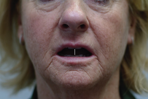
Figure 3.20 Mouth slightly open, displaying alignment of dental midlines (white solid line).
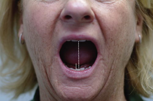
Figure 3.21 Maximal unassisted opening, with mandible deviated to the patient’s right. Note solid white line positioned at dental midline, relative to the dashed reference line in the mid-sagittal plane extending from the incisal embrasure of the maxillary central incisors.
Neck
- Cervical mobility is normal for flexion, extension, bilateral rotation, and bilateral side-bend.
G. Intraoral Status
Soft tissues and hard tissues
- Unremarkable.
Occlusion
- Unremarkable.
Saliva
- Not evaluated.
H. Additional Examinations and Findings
Physical therapy consultation was requested in order to assess for any other treatment options. Evaluation revealed the following:
- Normal seated and standing postural curves. Negative cervical spine segmental screen for somatic dysfunction.
- Symmetrical hypertonic cervical extensors with full cervical spine active and passive range of motion.
- Palpation pain present in the submandibular muscles on the left, as examined intraorally, adjacent to the prior tooth extraction site, and in the symmetrical cervical extensors. Palpation pain and tightness absent in the sublingual muscles. Palpation tightness and pain in the medial aspect of the bilateral masseter muscles.
- Inferior and anterior glide of the TMJ resistant to passive mobilization on the right side.
- Initial maneuvers of grade 1–3 inferior and inferior–anterior mandibular glides until accessory motion was restored resulted in short-term reduction in resistance to the mobilizations, with gain of 2–3 mm in jaw opening with less deviation, demonstrating viability of possible improvement in opening.
I. Diagnosis/Diagnoses
Expanded DC/TMD
- Bilateral masseter contracture.
Other
- Right TMJ capsular contracture.
J. Case Assessment
- The contracture of the masseter muscles and right TMJ capsule are secondary to prolonged limitation in opening which was a consequence of the TMJ mechanical disc problems associated with an motor vehicle collision 35 years previously and not addressed at that time. Because of the 3 year period during which the mechanical disc problems continued, loss of normal resting length of the masseter muscles would have likely occurred; the limitation in opening due to contracture versus the limitation in opening due to the mechanical disc problems can be very challenging to distinguish, and given the history of the whiplash trauma (see Case 3.10) and the clear history of mechanical disc problems, treatment was focused primarily on the disc problems, and the contracture was not addressed.
- The left TMJ sounds at present meet the criteria for disc displacement with reduction; however, those sounds are not relevant in terms of chief complaint or functional status, given the absence of mechanical locking, and therefore this diagnosis is set aside and further investigation via MRI is not recommended.
- The contracture is presently stable but is also responsive to guarding behaviors, evident on examination and supported by history, resulting in nonsymmetrical restriction on opening.
- History of recent trismus was secondary to a dental treatment visit; possible causes include injection trauma by the anesthesia, prolonged mouth opening, force on the mandible during extraction, and exacerbation of factors associated with perpetuating the chronic contracture. The relatively fast recovery was in part due to her self-management skills and determination to return to her prior level of functioning.
- Medically and psychosocially, she is doing extremely well, and there are no identified major risk factors at this time for active contribution to the contracture; therefore, the contracture is regarded as stable.
- While the patient would like to open her mouth wider, she is realistic and would rather remain with the current restrictions and mild symptoms that she effectively self-manages than risk aggravating the condition with new treatment that may not yield substantial benefits.
K. Evidence-based Treatment Plan including Aims
- Provide reassurance regarding the temporary state of the recent trismus and the continued stability of the contracture in the absence of any further treatment.
- Describe, as part of patient education, possible treatment as follows, recognizing that the model for care is based on that which has been developed for adhesive capsulitis of the shoulder (see later):
- Manual therapy to promote elasticity in contracted capsule and ligaments.
- Massage to hypertonic muscles to prepare for movement reeducation.
- Postural reeducation to promote balanced structures and optimal muscle length and strength.
- Exercise for reeducation of functional jaw movement.
- Stretching to tolerance to retain or gain motion.
L. Prognosis and Discussion
- Patient has adapted to limited opening due to chronic contractures and she experienced a loss of jaw opening following a recent dental procedure; it is likely that the chronic contracture contributed to the development of the trismus, first via restricted opening that hindered normal dental procedures (e.g., more strain was placed on the mandible for sufficient intraoral access), and second via loss of normal soft tissue elasticity, such that the tissues did not recover normally from any prolonged stretch during the dental procedure.
- Long-term prognosis remains favorable for retaining a functional opening and restoring any remaining lost mobility following the recent dental procedures.
- Self-management strategies have been effective and include gradual stretching and awareness of posture, and maintaining cervical spine mobility and elasticity of masticatory and cervical soft tissues.
Background Information
- The most dramatic form of musculoskeletal contracture depicts arms and legs unable to straighten, stuck in bent angles with minimal hope of returning to normal. In contrast, we focus here on musculoskeletal contracture that involves the abnormal shortening of muscle and which may also include the associated tendons as well as joint ligaments and capsule. Most such contractures are not dramatic but rather are often inadequately identified.
- Contractures are most commonly associated with prior trauma and prolonged immobilization. One of the most common non-trauma etiologies, and for which good data exist, is adhesive capsulitis of the shoulder (“frozen shoulder”), which has estimated prevalence of 2–5% of the general population (Reeves, 1975). Masticatory system contractures probably occur at a much lower rate.
- Loss of mobility within the masticatory system can occur without rapid detection because most individuals have greater jaw mobility than required for typical functional demands of food ingestion; by one study, 95% of the US adult population exhibit a maximal nonassisted jaw opening of 39 mm or greater, more than sufficient for food ingestion (Ohrbach et al., 2011).
- An initial loss of jaw mobility is readily compensated by adaptive movements permitted by the bilateral joint system. Because initial mobility losses are often undetected, an insidious development of contractures in muscle and associated connective tissues may occur. Within the masticatory muscles, the prevalence of contracture is unknown but believed to be much lower than the prevalence of masticatory muscle myalgia, for example. Challenges in both diagnosis and treatment accompany this condition.
- There are no published data regarding demonstrated etiologic pathways for masticatory muscle contracture. Extrapolating from other joints for which mechanisms have been established, the available evidence suggests the following as contracture mechanisms of the masticatory system: altered biomechanics likely predispose the TMJ to develop contractures, and altered mandibular posture (a deviation from anatomic neutral which alters muscle length–strength relationships) likely sets the stage for contractures.
- If the primary mechanism of contracture in the jaw targets the masticatory muscles, then such contractures most certainly lead to altered biomechanics of the TMJ, thereby establishing a positive feedback loop leading to yet further worsening of a muscle contracture.
- Time is the major factor affecting contracture severity: the longer a muscle or joint tissue is maintained in a shortened position, the more likely that reduced mobility will become permanent and have repercussions throughout that system and adjacent musculoskeletal system.
Stay updated, free dental videos. Join our Telegram channel
VIDEdental - Online dental courses


